
On 9 October 1676, a cloth merchant in Delft, in the Netherlands, wrote a long letter to the Royal Society in London. He had soaked peppercorns in water for three weeks, put a drop of it in front of a tiny glass bead, and seen “very little animalcules”, so small that, he guessed, a hundred of them laid end to end would not reach the length of a grain of sand. He was the first person ever to see bacteria. Today we know they are the most numerous living things on Earth: a pinch of soil from a Bhaktapur field holds more of them than there are people in Nepal.
This unit, the first of microbiology, the study of living things too small to see with the naked eye, is about them: the kingdom Monera, the bacteria and the cyanobacteria.

3.1.1 General introduction
The kingdom Monera includes the most ancient, smallest and simplest forms of life. All prokaryotes belong to it. Prokaryote comes from Greek pro, before, and karyon, kernel or nucleus: organisms whose cells lack a true nucleus. They live everywhere on Earth: in hot springs, in water saturated with salt, inside other living things, deep inside the Earth’s crust and at the bottom of the ocean. Bacteria were probably the earliest forms of life: fossil traces of their mats are about 3.5 billion years old. A teaspoonful of fertile soil may contain hundreds of millions of bacteria.
The organisms of this kingdom are:
- microscopic and unicellular (single cells, though some live in chains, filaments or colonies);
- prokaryotic: the cell has no organized nucleus and no membrane-bound organelles (no mitochondria, chloroplasts, Golgi bodies or endoplasmic reticulum);
- covered by a rigid cell wall;
- with circular, naked DNA as their genetic material, lying in the cytoplasm. Naked means it is not wrapped round histone proteins as eukaryotic DNA is. This DNA, with no membrane round it, is the cell’s only “nucleus”, and is called the nucleoid (incipient nucleus);
- autotrophic (making their own food) or heterotrophic (taking it from others), and the major decomposers of every ecosystem;
- without true sexual reproduction: there is no fusion of gametes. (Bacteria can still swap genes, by the ways described under 3.1.2, so they are not simply copies of their parents.)
The kingdom Monera is divided into two groups:
3.1.2 Bacteria
Bacteria are the simplest, smallest unicellular prokaryotes, able to survive in almost every kind of environment: wherever life can exist, bacteria are found. They were first seen by the Dutch naturalist Antonie van Leeuwenhoek in 1676. The branch of biology that deals with bacteria is bacteriology.
Bacteria are the smallest of all known cellular organisms. Most are about 0.5 µm to 1.5 µm across (a micrometer, µm, is a thousandth of a millimeter), and a few micrometers long. So about 500 of them would fit side by side across the full stop at the end of this sentence.
Most bacteria keep the same shape throughout their lives, and it helps to identify them. Some, though, change their shape and size when conditions change: this ability is pleomorphism, as in Corynebacterium (the cause of diphtheria) and Legionella pneumophila.
Shapes and forms of bacteria
On the basis of shape, bacteria are of four kinds.
1. Cocci (singular coccus; Greek kokkos, berry). Spherical, oval or round. Most cocci have no flagella, so they can’t swim. After dividing, the daughter cells may stay together in a pattern, which depends on how the cells divide:
- monococcus: single cells;
- diplococcus: in pairs (Diplococcus pneumoniae, now Streptococcus pneumoniae, a cause of pneumonia);
- tetracoccus: in groups of four, from division in two planes;
- streptococcus: in chains, from division always in one plane (Streptococcus thermophilus in curd; Streptococcus pyogenes in sore throats);
- staphylococcus: in irregular clusters like a bunch of grapes, from division in random planes (Staphylococcus aureus, boils and wound infections);
- sarcina: in cubes of eight or more, from division in three planes at right angles.
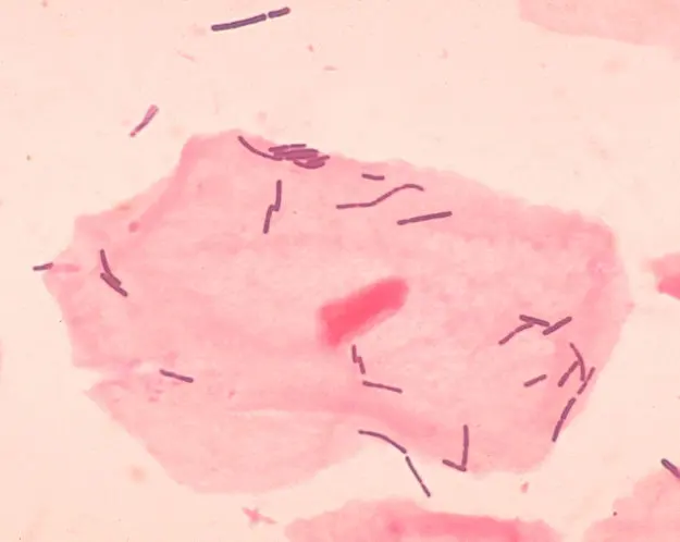
2. Bacilli (singular bacillus; Latin, little rod). Rod-shaped or cylindrical. Some bear flagella. Their forms:
- bacillus: single rods (Escherichia coli);
- diplobacillus: rods in pairs;
- streptobacillus: rods in chains (Bacillus anthracis, anthrax);
- palisade: rods lying side by side like a stack of matches or a fence (palisade), as in Corynebacterium diphtheriae.
3. Helical (spiral) bacteria. Curved or coiled:
- vibrio: shaped like a comma, a short curve (Vibrio cholerae, cholera);
- spirillum: a rigid spiral of several turns (Spirillum volutans). (Thin, flexible spirals that wriggle, such as Treponema, the cause of syphilis, are called spirochetes.)
4. Filamentous bacteria. Long, thread-like filaments of cells, often branched, like the fungus-like Streptomyces of the soil, which gives fresh earth its smell after the first monsoon rain and gives us the antibiotic streptomycin.
Mariana Ruiz Villarreal, LadyofHats, Public domain, via Wikimedia Commons
Under the microscope, a sample shows round cells in long, bead-like chains. What are they?
Pick an answer to see why.
Structure of a bacterial cell
A bacterial cell is simple: it has no organized nucleus and no membrane-bound organelles such as mitochondria, Golgi bodies or plastids. From the outside in:
a. Slime layer and capsule. Many bacteria cover their wall with a sticky, jelly-like layer. A thin, loose one is a slime layer; a thick, firm one is a capsule, and a bacterium with one is capsulated. Both are made mostly of polysaccharides (sugars; in a few bacteria, such as the anthrax bacillus, of protein). They protect the cell from drying out (desiccation), from chemicals, and in a host from being swallowed by white blood cells; they also help bacteria stick to surfaces, such as the plaque on your teeth.
b. Cell wall. A rigid protective covering made of peptidoglycan (also called murein or mucopeptide), a mesh of sugars cross-linked by short chains of amino acids. It gives the cell its shape and stops it bursting when water flows in; it protects it from mechanical injury. (Penicillin works by stopping bacteria from building this wall.)
c. Plasma (cell) membrane. Inside the wall, a thin, elastic, selectively permeable membrane of lipids and proteins, which controls what enters and leaves. Since bacteria have no mitochondria, the enzymes of respiration are on the inner surface of this membrane: the cell membrane is the site of respiration in bacteria.
d. Cytoplasm. The granular, colorless fluid inside the membrane. In it are:
- ribosomes of the 70S type (smaller than the 80S ribosomes of eukaryotes), where proteins are made;
- reserve food granules of glycogen, lipid or protein;
- gas vacuoles in some aquatic bacteria, which help them float;
- in photosynthetic bacteria, folds or sacs of membrane, the chromatophores (thylakoids), which carry the photosynthetic pigments.
e. Nucleoid. A single, circular, double-stranded, naked DNA molecule, folded and twisted into a tangle near the center of the cell, not enclosed by any membrane.
f. Plasmids. Many bacteria also have small rings of extra DNA, separate from the main chromosome: extrachromosomal genetic elements. They copy themselves on their own and often carry genes that help in hard times, such as resistance to antibiotics, and they can pass from one bacterium to another.
g. Flagella (singular flagellum). Long, fine, whip-like threads of protein growing from the cell, which turn like propellers to make the bacterium swim (motile bacteria). A capsulated bacterium can have flagella too: they grow out through the capsule, as in E. coli and Salmonella.
h. Pili (singular pilus; also fimbriae). Short, fine, hair-like threads on the surface, more numerous than flagella, which help bacteria attach to surfaces and to a host’s cells; a special, longer sex pilus joins two bacteria during conjugation.
Mariana Ruiz Villarreal, LadyofHats, Public domain, via Wikimedia Commons
A bacterium has its DNA as a circular molecule without histones, free in the cytoplasm. What is this DNA called?
Pick an answer to see why.
3.1.3 Mode of nutrition in bacteria
Like all living things, bacteria need nutrients and energy to grow. They get them in two main ways: autotrophic (making their own food) and heterotrophic (taking it from other organisms).
1. Autotrophic bacteria
A small number of bacteria are autotrophic: they make their own organic food from inorganic substances (carbon dioxide and water or other simple compounds), using sunlight or chemical energy. There are two kinds.
a. Photoautotrophic bacteria use light energy to make food by photosynthesis. But their photosynthesis differs from that of green plants: they don’t use water as the source of hydrogen (the hydrogen donor), so no oxygen is given off. This is anoxygenic photosynthesis. Instead of water, they take hydrogen from hydrogen sulphide (H₂S), thiosulphate, hydrogen gas, or organic compounds. The green sulphur bacteria (Chlorobium) and purple sulphur bacteria (Chromatium), for example, live in sulphur springs and the mud of ponds and use H₂S, giving off sulphur instead of oxygen:
Compare the green plant’s equation, with water in place of H₂S and oxygen in place of sulphur. (Cyanobacteria are the exception among bacteria: they split water and give off oxygen, as plants do; see 3.1.5.)
b. Chemoautotrophic bacteria use chemical energy, released by oxidizing simple inorganic compounds, to turn carbon dioxide into organic food: this is chemosynthesis. They need no light. By the compound they oxidize, they are:
| Group | Oxidizes | Example |
|---|---|---|
| nitrifying bacteria | ammonia to nitrite, or nitrite to nitrate | Nitrosomonas, Nitrobacter |
| sulphur bacteria | hydrogen sulphide (or sulphur) | Beggiatoa, Thiobacillus |
| iron bacteria | ferrous (iron II) compounds to ferric | Gallionella, Ferrobacillus |
| hydrogen bacteria | hydrogen gas | Hydrogenomonas |
The methane-oxidizing bacteria (Methylococcus), which use methane, are often listed with them. Methane, though, is an organic compound, and they take their carbon from it, so strictly they are not autotrophs.
2. Heterotrophic bacteria
Heterotrophic bacteria can’t make their own organic food; they depend on other organisms. Most bacteria are heterotrophic. There are three kinds.
a. Saprophytic bacteria feed on dead and decaying organic matter. They secrete digestive enzymes onto it, which break complex organic matter (proteins, fats, cellulose) into simpler forms, and absorb the products. In doing so they release nutrients back into the soil and air for plants to use again: recycling of nutrients by decomposition. So they are the decomposers of an ecosystem, nature’s scavengers. Examples: Pseudomonas, Acetobacter (which turns alcohol into vinegar), Clostridium, and the Bacillus that rots fallen leaves.
b. Parasitic bacteria get their food from the living cells of other organisms (the host), usually harming it. Those that cause disease are pathogenic: Mycobacterium tuberculosis (tuberculosis), Clostridium tetani (tetanus), Salmonella Typhi (typhoid), Vibrio cholerae (cholera).
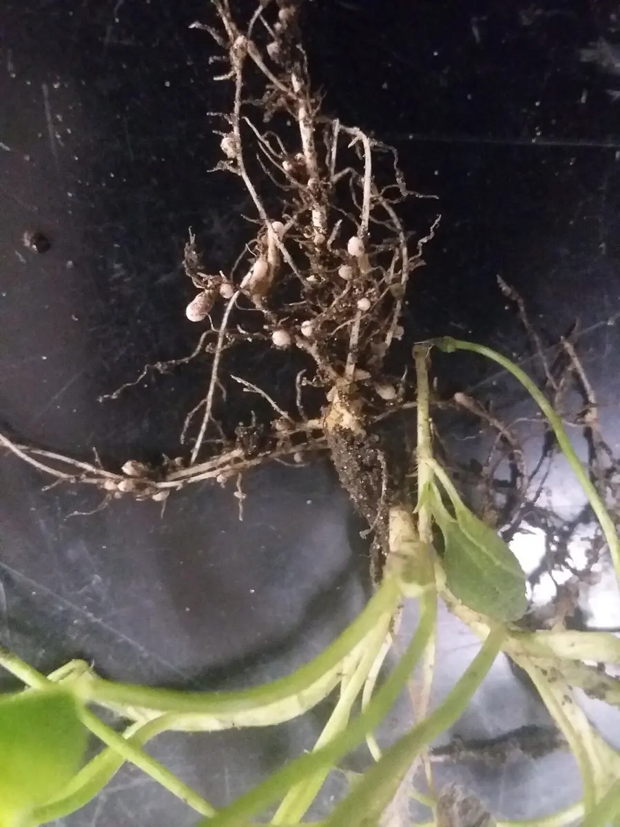
c. Symbiotic bacteria live in a close association with another organism in which both partners benefit (mutualism). Rhizobium lives in the root nodules of legumes (pea, bean, gram, soybean): it fixes nitrogen from the air into ammonia, which the plant uses to make proteins, and the plant gives it sugars and a safe home. Escherichia coli in the human large intestine is usually harmless and makes vitamin K, which we absorb; in return it gets food and warmth. The bacteria in the rumen of a cow or buffalo digest the grass’s cellulose for it.
A bacterium living in the mud of a dark ditch takes in carbon dioxide and makes sugar, getting its energy by oxidizing ammonia. How does it feed?
Pick an answer to see why.
3.1.4 Bacterial growth
In plants and animals, growth means an increase in size. In bacteria it means an increase in number. Bacteria grow by binary fission, a form of asexual reproduction in which one cell copies its DNA, lengthens, and divides across the middle into two cells of the same size.
Mariana Ruiz Villarreal, LadyofHats, Public domain, via Wikimedia Commons
Each division doubles the number, so a bacterial population grows in a geometric or exponential way: 1, 2, 4, 8, 16, 32, 64… After generations, one cell has become cells. The time taken for one doubling is the generation time: about 20 minutes for E. coli in warm broth, about 15–20 hours for the slow tuberculosis bacterium.
Bacteria need the right conditions to grow, and these are not the same for all of them: oxygen (some need it, some are killed by it, some can do either), pH (most prefer near neutral), temperature (most disease-causing bacteria grow best at our body temperature, 37 °C; others in hot springs above 80 °C), light, osmotic pressure (salt and sugar draw water out of them, which is why pickles keep), atmospheric pressure, the moisture available, and the nutrients in their surroundings.
The growth curve
When a nutrient broth (a culture medium) is inoculated with a few bacteria and kept warm (incubated) for long enough, the number of living bacteria follows a typical pattern. A graph of the number of living cells in a population against time is the bacterial growth curve. Because the numbers grow so fast, it is drawn with the number on a logarithmic scale (each step up the scale is ten times the one below), so that steady doubling shows as a straight line. It has four phases:
- Lag phase. The bacteria have just been put into a new medium. They are metabolically active but not dividing: they adjust to the new surroundings and prepare to divide, making the enzymes and other molecules they need. The number stays nearly the same.
- Log phase (exponential phase). The cells divide continuously at their fastest steady rate, doubling every generation time: the number increases exponentially, a straight line on the log scale. The cells are at their most active, and most uniform; antibiotics such as penicillin, which act on growing cells, work best now.
- Stationary phase. The food is running out and toxic waste products made during the log phase (acids, for example) are building up. The number of dividing cells equals the number dying, so there is no net increase in the population: the curve is flat.
- Decline phase (death phase). As the food runs out and the waste builds up further, the number of dying cells exceeds the number of new cells, and the population falls.
During which phase of the growth curve is the number of new cells equal to the number of cells dying?
Pick an answer to see why.
3.1.5 Cyanobacteria
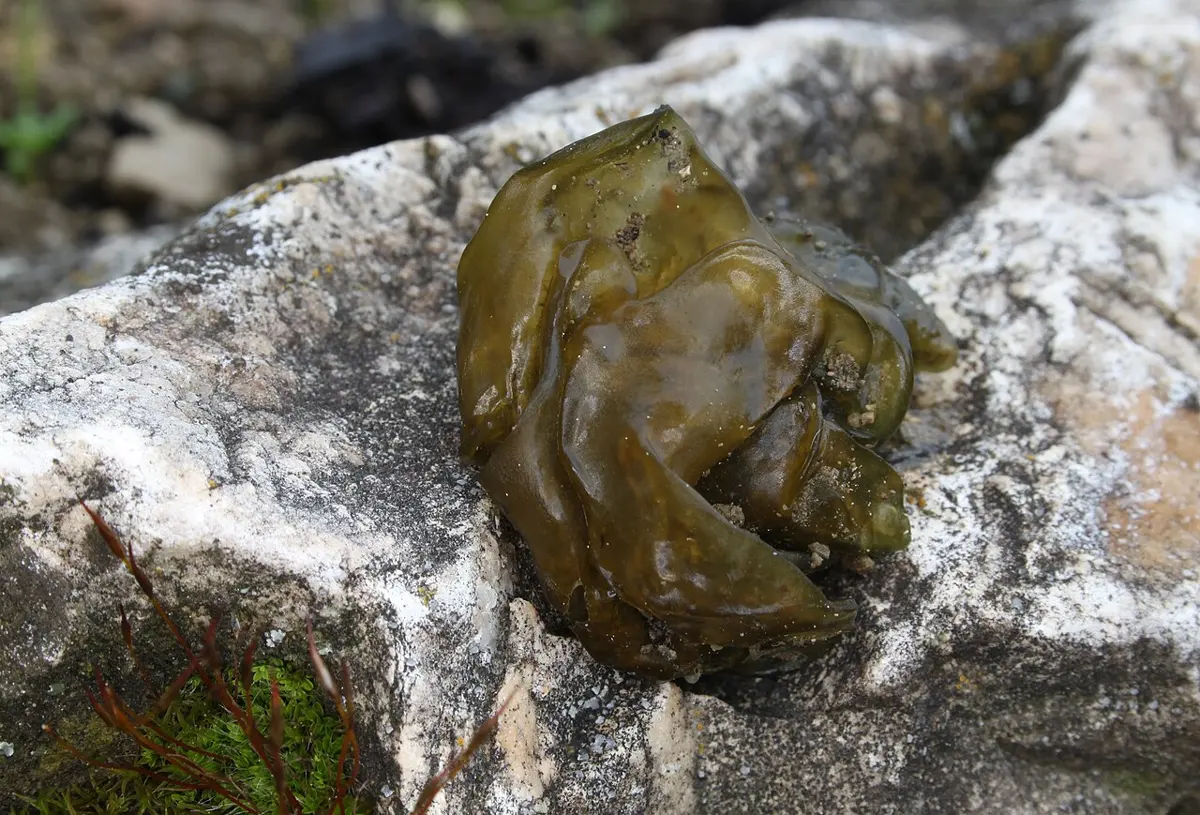
Cyanobacteria (Greek kyanos, dark blue), once called blue-green algae, are photosynthetic bacteria, found in fresh water and the sea, in moist soil, on damp walls, tree bark and rocks, even in hot springs and on glaciers. They live singly or in colonies. They are among the oldest forms of life on Earth: layered mounds built by their mats, stromatolites, are found as fossils over 3 billion years old, and still grow today in a few warm, salty bays in Australia.
Most contain the green pigment chlorophyll a (with other pigments), which traps the Sun’s energy, so they are autotrophic. Unlike other photosynthetic bacteria, they split water and give off oxygen, as plants do. Some live in symbiosis with other organisms: with fungi in lichens, and Anabaena inside the leaves of the water fern Azolla.
Features of cyanobacteria:
- a.They may be unicellular (Chroococcus, Synechococcus) or multicellular: filamentous (Nostoc, Anabaena, Oscillatoria) or colonial (Microcystis).
- b.Each cell is surrounded by a rigid cell wall (of peptidoglycan, like other bacteria), covered by a mucilaginous sheath; inside the wall is a plasma membrane of lipids and proteins.
- c.The cell lacks membrane-bound organelles such as mitochondria, Golgi bodies and chloroplasts.
- d.Their photosynthetic pigments are chlorophyll a, carotenes, xanthophylls and the phycobilins: blue phycocyanin and red phycoerythrin, which give them their blue-green (sometimes red or brown) color. The pigments lie on flattened sacs of membrane, the thylakoids, in the outer part of the cytoplasm (the chromoplasm); there are no chloroplasts.
- e.In the center of the cell is the nucleoid (the centroplasm), with circular DNA without histones.
- f.They have no flagella. (Many filaments, such as Oscillatoria, still glide slowly over surfaces.)
- g.In a filamentous cyanobacterium, the row of cells is the trichome; the trichome with the slimy sheath round it is the filament. In Nostoc, many twisted filaments lie together in a jelly-like ball (a colony).
- h.Some filamentous ones (Nostoc, Anabaena, Scytonema) have special, larger, thick-walled, pale cells in their filaments, the heterocysts, where nitrogen fixation takes place. (The enzyme that fixes nitrogen, nitrogenase, is destroyed by oxygen; a heterocyst stops making oxygen and shuts it out.)
- i.Their cells divide by binary fission, as in all bacteria (there is no mitosis, which needs a nucleus and a spindle). They reproduce by vegetative methods (fission; the filament breaking at the heterocysts into pieces called hormogonia) and asexual methods (thick-walled resting spores, akinetes). Sexual reproduction is absent.
Nostoc: L. H. Pammel, A Manual of Poisonous Plants (1910), public domain
Where does nitrogen fixation take place in Nostoc?
Pick an answer to see why.
Exercise
A. Choose the correct option
- Name the site of respiration in bacteria.
Pick an answer to see why.
- Which of the following genomes is present in bacteria?
Pick an answer to see why.
- Which one of the following is absent in bacteria?
Pick an answer to see why.
- At which phase of bacterial growth does the rate of dying of cells exceed the rate of new cells being formed?
Pick an answer to see why.
- Which of the following statements describes the characters of the kingdom Monera?
Pick an answer to see why.
- Agricultural scientists want to improve crop yield and soil health by introducing nitrogen-fixing bacteria into the soil. Which crop would be the most suitable, considering its ability to form nodules with nitrogen-fixing bacteria?
Pick an answer to see why.
- How do bacteria contribute to nutrient cycling and waste decomposition in an ecosystem?
Pick an answer to see why.
B. Answer the following questions
Define plasmid.
Answer
What do you mean by naked DNA?
Answer
What types of bacteria are known as symbiotic bacteria? Give examples.
Answer
Compare the characteristic features of autotrophic and heterotrophic bacteria.
Answer
Explain the structural features of a bacterial cell, with a labeled diagram.
Answer
Explain why blue-green algae are classified in the kingdom Monera rather than the kingdom Plantae. Include specific characteristics of blue-green algae, and compare them with both plants and bacteria to justify their placement.
Answer
Study the bacterial growth curve above and answer:
- a.Which phase of the bacterial growth curve has rapid cell division?
- b.What happens during the lag phase?
- c.In which phase is there a balance between cell division and cell death?
- d.Why do the dying cells outnumber the newly formed cells during the death phase?
Answer
Many organisms die, but their bodies do not remain as they are. Where do their bodies go? Do you think any organisms have a role in making their bodies disappear?
Answer
Describe the ecological roles and significance of organisms classified in the kingdom Monera, with examples of their impact on ecosystems and human activities.
Answer
Printable PDFs
- Study notes 3 pages
- Worksheet 5 pages Answer key
Quiz yourself: Classification of Living Beings, Grades 5–12 →
Practice every question in the book, and keep track of the ones you miss →
A companion to your textbook, not a replacement for it: study from the book your school uses, and where the two differ, follow your teacher. Found a mistake? Please tell us.