
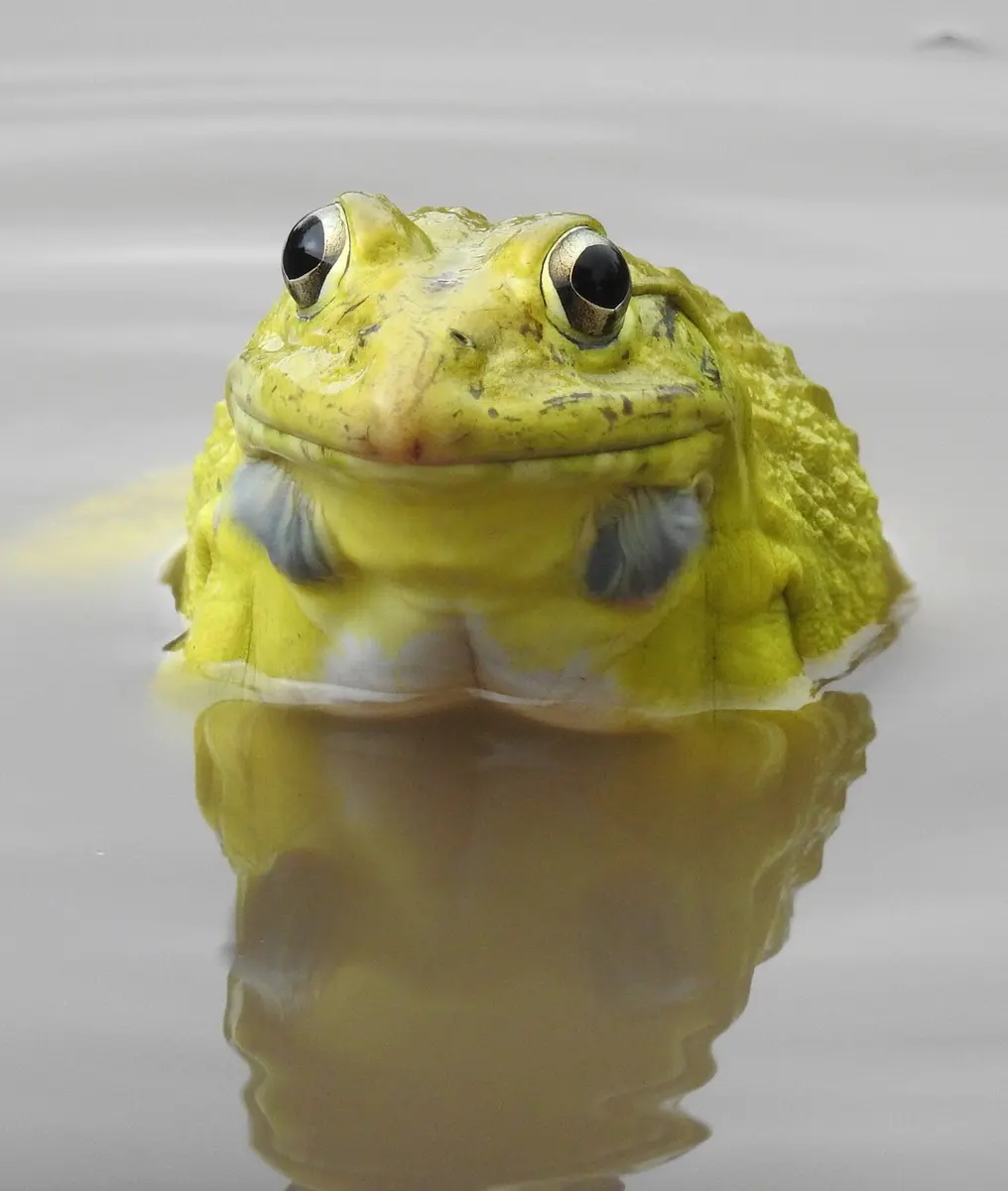
On a monsoon night in the Terai, every flooded paddy field seems to roar. The sound comes from frogs, and the loudest are big bright-yellow males, each with a pair of blue balloons blowing out from the corners of his mouth. A few weeks later the same water is thick with tadpoles, and by autumn the frogs have gone quiet, ready to dig into the mud and sleep through the cold.
This unit looks at one of those frogs, the Indian bullfrog, from its skin inwards: how it catches and digests an insect it never chews, how one heart with a single pumping chamber sends blood to its lungs, skin and brain, how it breathes with its mouth, skin and lungs all at once, and how a new generation begins in a pond.
Observe and discuss
8.4.1 General introduction
Systematic position
| Rank | Name |
|---|---|
| Kingdom | Animalia |
| Phylum | Chordata |
| Group | Craniata |
| Subphylum | Vertebrata |
| Division | Gnathostomata |
| Superclass | Tetrapoda |
| Class | Amphibia |
| Genus | Rana (now Hoplobatrachus) |
| Species | tigrina (now tigerinus) |
| Common name | Indian bullfrog |
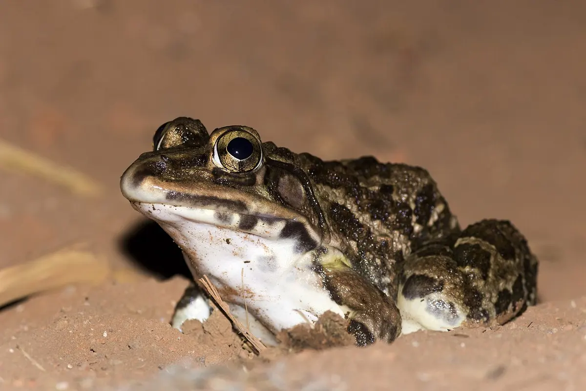
School books have long called this frog Rana tigrina. Zoologists have since moved it to its own genus, and its scientific name is now Hoplobatrachus tigerinus. Its common names are the Indian bullfrog or Asian bullfrog. It lives in the lowlands of southern Asia, from Afghanistan and Pakistan through India, Nepal and Bangladesh to Myanmar; in Nepal it is the big frog of the Terai’s ponds, ditches and paddy fields.
It is easy to tell from other frogs: it is large; it is olive-green to brown above with dark blotches; its snout is pointed; a pale line often runs down the middle of its back; and the breeding male has a pair of vocal sacs for croaking. It lives in moist places, forests, farmland and even towns, but always near fresh water: ponds, lakes, rivers and flooded fields.
Frogs are cold-blooded (poikilothermic): their body temperature follows the air and water around them. So they hide in moist burrows through the coldest days of winter and the hottest days of summer. People in some countries farm or hunt frogs for their meat. Frogs are natural predators of insects that damage crops, and they have been used in medical and scientific research for centuries, because in their body plan and working they are surprisingly like other vertebrates, ourselves included.
Why the frog is chosen for study
- i.It is easy to find, cheap and harmless (not poisonous).
- ii.The bullfrog is much better known than the toad.
- iii.It is a good size to handle and dissect in a laboratory.
- iv.It can be kept alive for a few days without feeding.
- v.Its life cycle is short and simple, and easy to watch.
- vi.It is a good sample of the vertebrates: its anatomy is built on the same plan as that of higher vertebrates, including humans.
8.4.2 Habitat and habit
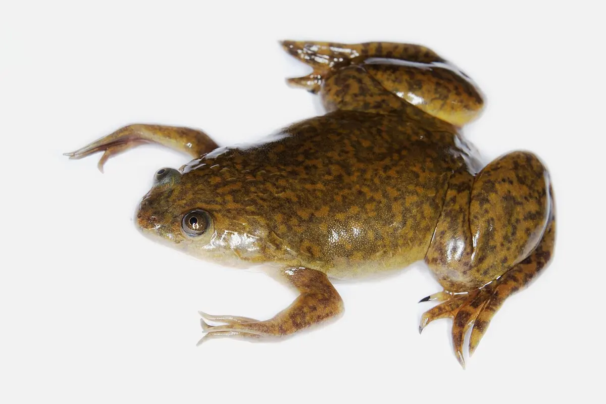
The bullfrog lives in water or on damp ground. Most of the time it sits on moist land with plenty of green plants near fresh water, or swims in ponds, ditches, rivers, lakes and streams. Living near water suits it in four ways:
- it visits the water often to keep its skin moist for breathing through the skin (cutaneous respiration);
- it escapes from enemies by diving straight into the water;
- its food, insects, worms, slugs and small frogs, is plentiful there;
- it needs water to breed: its eggs, embryos and tadpoles all live in water.
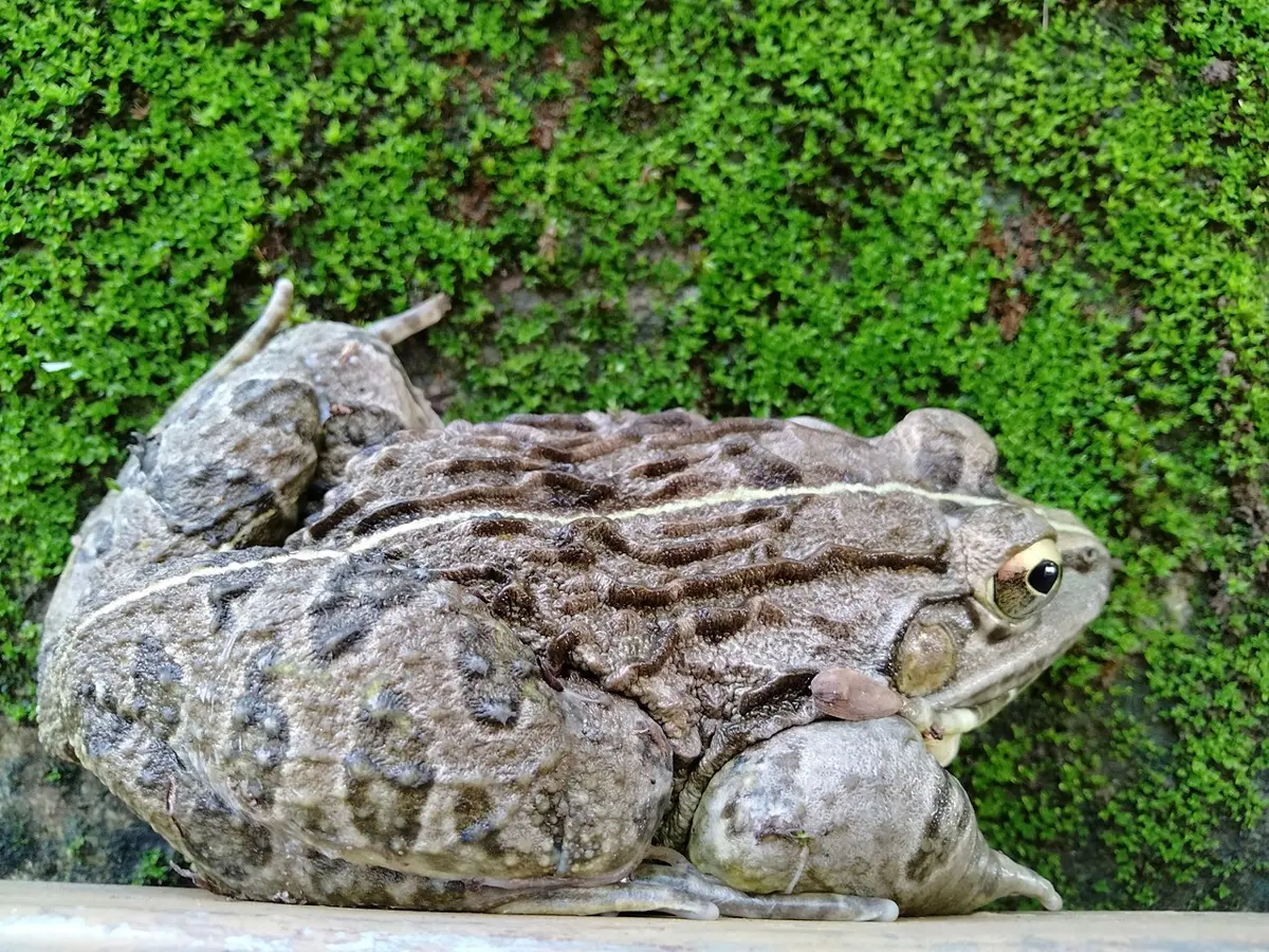
The bullfrog is harmless, solitary and usually silent, with no weapons such as claws or poison. It is free-living and a predator, and it is cannibalistic: a big frog eats smaller frogs. Usually you notice it only when you disturb it and it leaps into a pond, a stream or a paddy field. It is mostly nocturnal: it rests in cover near the water by day and hunts at dusk and at night. In the breeding season the males gather in shallow water in paddy fields and ponds and croak.
i. Locomotion
On land a frog leaps; in water it swims. At rest it sits with its long, strong hind legs folded in a Z under the body and its short front legs held upright. To leap, it straightens its hind legs suddenly, throwing the body forwards into the air, and it lands on its front legs, which take the shock. A big bullfrog can leap more than a meter. In water, the hind legs kick backwards together, the broad webbed feet spreading to push against the water; the front legs help with steering.
ii. Feeding
The adult frog is carnivorous. It eats living insects, worms, slugs, small fish, tadpoles and small frogs. It flicks out its long, sticky tongue, catches the prey and pulls it in, then swallows it whole, without chewing. It hunts only moving prey and ignores still or dead animals. Before winter it eats greedily and stores glycogen and fat in its body, the reserve it lives on during hibernation.
A frog flicks out its tongue at a beetle. Which describes what happens next?
Pick an answer to see why.
iii. Hibernation and aestivation
Frogs are poikilothermic (cold-blooded): their body temperature changes with their surroundings. In a cold winter it could fall low enough to kill them. So they hide, inactive, in moist burrows or in the soft mud at the bottom of ponds. This winter rest is hibernation, or “winter sleep”, and it lasts through the coldest months. During hibernation:
- the nostrils and mouth stay closed, and the frog stops feeding and excreting;
- its metabolism slows right down;
- it breathes only through its skin;
- it lives on the glycogen and fat stored in its body.
In the hottest, driest part of the summer, frogs rest in the same way, buried in mud or burrows, to escape the heat and drought. This is aestivation, or “summer sleep”. It may last days or weeks depending on the weather, and the frogs come out now and then to feed when it is cooler.
iv. Croaking
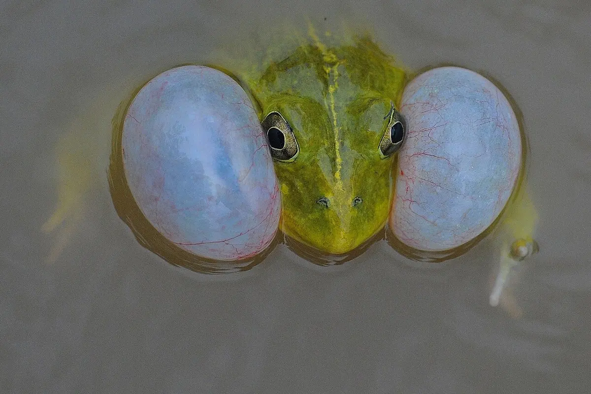
Croaking is the call of the male frog in the breeding season, a mating call that attracts females. Only males croak loudly. The breeding male has a pair of thin, stretchy skin pouches on the sides of his throat, the vocal sacs, which blow up with air like balloons and make the call louder.
v. Breeding
The rainy season (the monsoon, roughly June to September) is the breeding season. Breeding males turn a bright lemon yellow, gather in shallow water and croak to attract females. When a female comes, the male climbs on her back and grips her firmly round the body just behind her front legs, holding on with a rough, swollen pad on the first finger of each hand, the nuptial pad (copulatory pad). This embrace is called amplexus, or false copulation: there is no true copulation in frogs, and no sperm enters the female.
Amplexus stimulates the female to lay her eggs into the water through her cloaca. At the same moment the male pours his milt (seminal fluid with sperms) over them. So fertilization is external, in water. The mass of eggs held together in jelly is called spawn. The fertilized eggs (zygotes) develop and, in warm water, hatch within a few days into tadpoles, free-swimming and wholly aquatic larvae; after some weeks each tadpole metamorphoses into a small frog that can live on land and in water.
Frog stages: LadyofHats (CC0)
vi. Camouflage
A frog can change its color a little to match its surroundings, so that enemies do not notice it. Hiding by matching the background is camouflage. The color changes because of pigment cells in the dermis of the skin, the chromatophores: when the pigment inside them spreads out through the cell’s branches the skin darkens, and when it gathers into the middle of each cell the skin turns pale.
vii. Protection from enemies
The frog’s enemies are snakes, birds such as herons, cranes, hawks, eagles and crows, and people; bigger frogs eat smaller ones, and large numbers are killed for dissection. A frog protects itself in four ways:
- a.it changes its color to match its surroundings, so enemies can’t find it easily;
- b.it suddenly squirts urine when grabbed;
- c.its skin is slippery with mucus from the mucous glands, so it is hard to hold;
- d.it lives near water and dives in the moment an enemy comes.
A frog spends the winter buried in pond mud with its nostrils and mouth closed. How does it get oxygen?
Pick an answer to see why.
8.4.3 External features (morphology)
LadyofHats, CC0, via Wikimedia Commons
1. Shape, size and color
The adult frog’s body is roughly spindle-shaped, pointed in front and rounded behind, flattened from top to bottom (dorsoventrally) and bilaterally symmetrical: one plane down the middle cuts it into matching left and right halves. A big Indian bullfrog grows to about 17 cm from snout to vent.
The skin is thin, moist, slimy and smooth, with no scales and no hard covering. It is slippery because of its mucous glands. A frog never drinks: it takes in water through its skin, mostly through a thin patch on its belly and thighs that it presses against wet ground. The moist skin is also a breathing surface.
The back (dorsal side) is green to olive-brown with dark spots and blotches; the belly (ventral side) is pale yellow or white. A thin pale mid-dorsal line runs down the middle of the back from the tip of the snout to the hind end.
2. Division of the body
The adult’s body has two parts: the head and the trunk, which carries the limbs. There is no neck and no tail.
i. Head. The head is flat and roughly triangular, ending in a short snout.
- The mouth is a wide slit across the front of the snout.
- Just above it are two small openings, the external nostrils (external nares), used in breathing.
- The two eyes bulge from the top of the head, one on each side. Because there is no neck to turn, bulging eyes let the frog see in almost every direction. Each eye has a thick, colored upper eyelid that cannot move and a thin, half-transparent lower eyelid that can. A third eyelid, the transparent nictitating membrane, joined to the lower lid, sweeps over the eye. It protects the eye under water and keeps it moist on land.
- Between the eyes is a small pale brow spot, which marks the place of a lost third eye (the pineal eye).
- Behind each eye is a round, colored disc of skin, the tympanum (eardrum). It picks up sound; a frog has no external ear.
- A breeding male has two blue, wrinkled vocal sacs on the sides of the throat, which blow up when he croaks.
ii. Trunk. The trunk is flat and oval, joined straight to the head. When the frog sits, its back rises in a hump near the hind end, the sacral prominence, where the backbone meets the hip girdle. At the hind end, between the hind legs, is a small round opening, the cloacal aperture (vent), through which feces, urine and eggs or sperms all leave the body.
Two pairs of limbs grow from the sides of the trunk.
- Each forelimb, just behind the head, has an upper arm (brachium), a forearm (antebrachium), a wrist (carpus) and a hand (manus) with four fingers; the thumb is missing (only a tiny bony trace is left inside). The fingers have no web. In a breeding male, the base of the first finger swells into the nuptial pad (copulatory or amplexus pad), which grips the female.
- Each hind limb is much longer and stronger. It has a thigh, a shank, a long ankle (tarsus) and a large foot with five toes, joined by folds of skin, the web, used in swimming.
- Under the fingers and toes are small round pads, the subarticular tubercles, which take the shock of landing and help the frog grip.
A frog has no neck. Which feature makes up for this most?
Pick an answer to see why.
Sexual dimorphism
Sexual dimorphism means that the male and female of one species can be told apart by their outward features. In the bullfrog it is clearest in the breeding season. In a pair in amplexus, the male is the one on top.
| Male frog | Female frog |
|---|---|
| Usually smaller | Usually larger |
| Turns bright yellow in the breeding season | Stays olive or brown |
| Nuptial pad at the base of the first finger in the breeding season | No nuptial pad |
| A pair of vocal sacs; croaks loudly | No vocal sacs; does not croak |
| Body slimmer | Belly swollen with eggs in the breeding season |
Toad and frog
Toads and frogs are both amphibians of the same order, Anura (the tailless amphibians). Nepal’s commonest toad, seen hopping round lights on summer nights, is the common Asian toad, Duttaphrynus melanostictus (once Bufo melanostictus); Europe’s common toad is Bufo bufo.
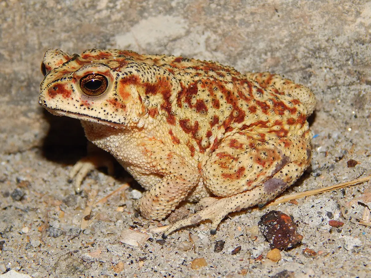
| Toad | Indian bullfrog |
|---|---|
| Mostly terrestrial; goes to water only to breed | Mostly aquatic; lives near water |
| Poisonous: a large parotoid gland behind each eye makes a poison | Not poisonous |
| Active at night (nocturnal) | Also mainly active at dusk and at night |
| Skin dry and rough, with warts | Skin moist, smooth and slippery |
| Snout short, broad and rounded | Snout narrower and pointed |
| Hind limbs shorter, toes only slightly webbed | Hind limbs longer, toes fully webbed: a good swimmer |
| Walks or makes short hops | Leaps on land and swims |
| No teeth in either jaw | Teeth on the upper jaw |
| Vocal sacs in the males of many toads (the common Asian toad has one; Europe’s common toad has none) | A pair of vocal sacs in the male |
| Lays eggs in long strings of jelly | Lays eggs in clusters (masses) |
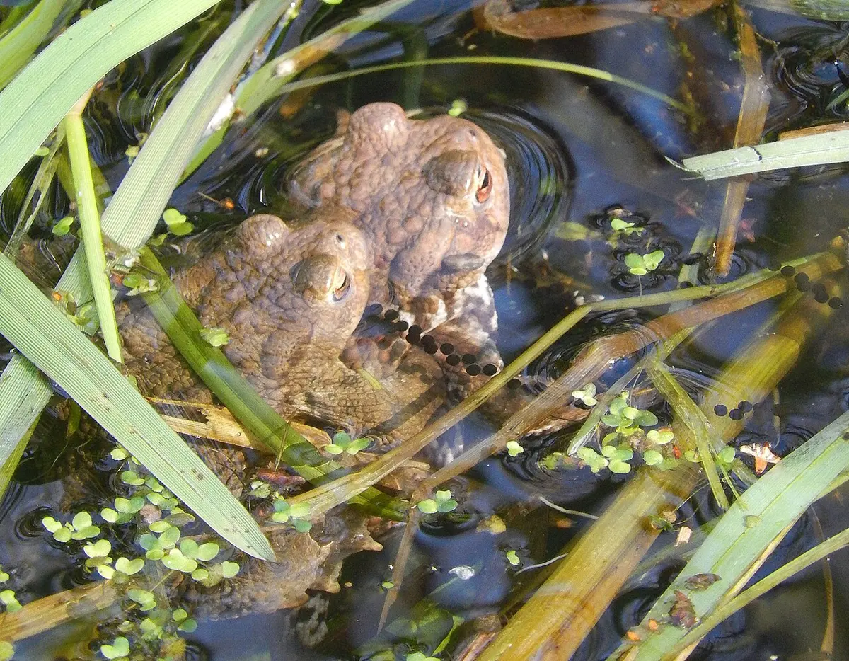
On a summer night you find an animal with dry, warty skin and a big swelling behind each eye, a long way from water. It is most likely:
Pick an answer to see why.
8.4.4 Digestive system
Digestion is the breaking down of large, complex food into simple, soluble substances that the body can absorb. It has two parts: mechanical digestion, which breaks big pieces into small ones, and chemical digestion, in which enzymes break complex molecules into simple ones. The organs that work together to do this make up the digestive system. In the frog it has two parts: the alimentary canal and the digestive glands.
A. Alimentary canal
The frog’s alimentary canal is a complete tube, long and coiled, of varying width, running from the mouth to the cloaca. In order: mouth → bucco-pharyngeal cavity → esophagus → stomach → small intestine (duodenum, ileum) → large intestine (rectum) → cloaca.
Alimentary canal: Gilchrist and von Bonde, Dissection of the Platana and the Frog (1919), public domain
1. Mouth
The canal starts with a wide opening, the mouth, a slit across the front of the head between the upper and lower jaws, edged by lips that cannot move. The upper jaw is fixed to the skull; the lower jaw moves up and down to close and open the mouth.
2. Bucco-pharyngeal cavity
The mouth opens into a wide space, the buccal cavity (mouth cavity). Because the frog has no neck, there is no clear line between this and the pharynx behind it, so the two together are called the bucco-pharyngeal cavity. It is the passage for both food and air. It is lined by ciliated columnar epithelium with mucus-making goblet cells; the mucus wets the food so it slides down. There are no salivary glands.
Mouth cavity: Gilchrist and von Bonde, Dissection of the Platana and the Frog (1919), public domain
The bucco-pharyngeal cavity contains:
i. Teeth. The upper jaw carries a row of small, sharp, conical teeth pointing backwards, set close together on the premaxillae and maxillae (the bones of the upper jaw): the premaxillary and maxillary teeth. The lower jaw has no teeth. Two more small patches of teeth stand in the roof of the mouth, one on each side of the middle line, on the vomer bones: the vomerine teeth.
Tooth: S. J. Holmes, The Biology of the Frog (1906), public domain
A frog’s teeth are not for chewing: they hold the prey after the tongue has caught it, so it cannot struggle out. They are:
- homodont: all of the same shape;
- pleurodont: fixed to the inner side of the jaw bone, leaning against it;
- polyphyodont: old and worn teeth fall out and new ones replace them again and again throughout life.
Each tooth is a cone with two parts, a broad bony base (root) joined to the jaw and a crown. The crown is mostly dentine, crossed by many fine branching canals, the canaliculi. Its tip is capped with enamel, a very hard, shiny white layer (made by cells called ameloblasts). Inside the tooth is the pulp cavity, filled with pulp, a soft tissue full of blood vessels and nerves, with dentine-making cells (odontoblasts) round its edge.
ii. Internal nostrils. In the roof of the mouth, beside the vomerine teeth, are two openings, the internal nostrils (internal nares, posterior nares), which lead to the external nostrils.
iii. Tongue. The tongue is large, thick, fleshy and protrusible (it can be thrown out). It lies on the floor of the mouth, fixed at the front to the inside of the lower jaw and free behind, where it is forked (bifid). Its upper surface has small papillae with taste buds. The secretions of tubular lingual glands and of the intermaxillary gland in the roof make it sticky. The frog flips it out over the front of the jaw, slaps it on its prey and pulls it back in a flash.
iv. Bulges of the orbits. Behind the vomerine teeth, the roof of the mouth has two large, soft, oval swellings: the undersides of the eye sockets and eyeballs. When the frog swallows, it pulls its eyes down into the head, and these bulges press the food backwards.
v. Eustachian openings. At the back corners of the roof are the openings of the two Eustachian tubes, which lead to the middle ears.
vi. Openings of the vocal sacs. A male frog has two openings in the floor of the mouth, one near each angle of the jaws, leading into the vocal sacs, which make his croak louder.
vii. Glottis and gullet. The glottis is a slit down the middle of the floor at the back, just behind the tongue. It opens to let air into the lungs and closes when the frog swallows. Behind it, the cavity leads into the esophagus through a wide opening across the back, the gullet.
viii. Fossae and prelingual elevations. The tip of the upper jaw has three small pits, the fossae, just inside the teeth; the tip of the lower jaw has three small bumps, the prelingual elevations. When the mouth closes, the bumps fit into the pits, sealing it tight.
A frog has caught a moth on its tongue. Which part of the mouth stops the moth struggling out while the frog swallows it?
Pick an answer to see why.
3. Esophagus
The bucco-pharyngeal cavity leads into the esophagus, a short, stretchy, muscular tube. Its lining is thrown into many lengthwise folds, which open out to let a large mouthful pass. It runs into the stomach with no clear boundary between them. Its lining has goblet cells and also glands that make pepsinogen.
4. Stomach
The stomach is a J-shaped, muscular bag on the left side of the body cavity, hung from the back wall by a fold of membrane, the mesogaster. Its front part, wide and long, is the cardiac stomach; its short, narrow hind part is the pyloric stomach. Its lining has many lengthwise folds, which flatten out when the stomach fills. The lining has:
- gastric glands, many-celled, that make pepsinogen;
- oxyntic cells, single cells that make hydrochloric acid;
- goblet cells, which make mucus.
The pyloric end is pinched in from outside, and its opening into the small intestine is guarded by a ring of muscle, the pyloric sphincter (pyloric valve), which stops food going back into the stomach. The stomach stores food for some hours and digests it in part.
5. Intestine
a. Small intestine. A long, narrow tube in several loops, held by a fan-shaped membrane, the mesentery. It is the main place of digestion and absorption. It has two parts.
- Duodenum: the first part, which turns forwards to make a U with the stomach. Its lining has low folds across it. The common bile duct (hepato-pancreatic duct), bringing bile from the liver and pancreatic juice from the pancreas, opens into it.
- Ileum: the rest, long, narrow and coiled. Its lining is raised into lengthwise folds; the frog has no true villi, and no crypts or glands like ours.
b. Large intestine. The ileum opens into a short, wide tube, the large intestine or rectum. Its lining has low lengthwise folds and goblet cells. It absorbs water and stores feces.
6. Cloacal chamber and cloaca
The rectum ends in a small bag, the cloacal chamber (cloaca), which also receives the ureters and, in the female, the oviducts. It opens to the outside through the cloacal aperture (vent) at the hind end of the body.
Which is the correct order of the frog’s alimentary canal?
Pick an answer to see why.
B. Digestive glands
Besides the small glands in the walls of the stomach and intestine, the frog has two large digestive glands: the liver and the pancreas.
1. Liver
As in other vertebrates, the liver is the largest gland of the body. It lies at the front of the body cavity, near the heart and lungs. It is reddish brown and has three lobes: right, middle and left, the left one split again in two. Between the right and left lobes, on the underside, lies the gall bladder, a round, thin-walled, greenish bag that stores bile. The liver cells make bile all the time; it reaches the gall bladder through the hepatic ducts. The cystic duct from the gall bladder joins some hepatic ducts to make the common bile duct, which runs through the pancreas, picks up the fine pancreatic ducts on the way, and opens into the duodenum. Bile has no digestive enzymes.
Under a microscope, the liver is made of many small lobules (hepatic acini) separated by connective tissue that carries bile capillaries, hepatic ducts, blood capillaries and sinusoids (wide blood spaces). Each lobule is a mass of many-sided gland cells, the hepatic cells, each with a nucleus and cytoplasm holding protein granules, fat droplets, glycogen and often black or brown pigment granules. The cells pour bile into fine channels, the bile canaliculi, which join into bile ductules and then the hepatic ducts. The liver gets blood from two vessels: the hepatic artery and the hepatic portal vein, which brings the blood from the gut.
Functions of the liver
- i.It makes bile, a green, alkaline juice of bile salts, bile pigments, cholesterol and water. Bile makes the food from the stomach less acidic, gives the pancreatic enzymes the alkaline medium they work in, and breaks large fat drops into tiny ones, emulsification.
- ii.It is the center of the metabolism of carbohydrates, proteins and fats. Extra glucose absorbed from the gut is turned into glycogen and stored (glycogenesis); when the body needs sugar, glycogen is turned back into glucose (glycogenolysis); and new glucose is made from things that are not carbohydrates, such as amino acids and glycerol (gluconeogenesis).
- iii.It does deamination and detoxification. Amino acids can’t be stored; the liver strips their amino groups off as ammonia, which is very poisonous, and joins the ammonia with carbon dioxide to make urea, much less poisonous, which the kidneys remove. (A tadpole, living in water, simply lets its ammonia out into the water; the frog switches to making urea as it changes into a land animal.)
- iv.It removes bilirubin and biliverdin, pigments from broken-down old red cells, in the bile, so they leave with the feces.
- v.In the tadpole the liver makes red blood cells; in the adult, it is one of the places where old red cells are destroyed.
- vi.Its Kupffer cells eat harmful microbes such as bacteria.
- vii.It stores iron, copper and the fat-soluble vitamins.
- viii.It makes fibrinogen and prothrombin, proteins needed for blood to clot.
2. Pancreas
The pancreas is a flat, irregularly branched, cream-colored gland held by the mesentery in the loop between the stomach and the duodenum. The common bile duct runs through it, and the pancreatic ducts open into this duct, making the hepato-pancreatic duct. The pancreas is a mixed gland: both exocrine and endocrine.
- Exocrine part. Under a microscope it is made of many small round groups of cells, the acini, set in connective tissue that also carries ducts, blood and lymph vessels and nerves. Each acinus is a ring of tall secretory cells, each with a large nucleus, round a small central space. The cells make pancreatic juice, with its enzymes, into this space; the spaces drain into ductules, the ductules join into the pancreatic ducts, and these open into the bile duct.
- Endocrine part. Scattered among the acini are small, compact groups of cells, the islets of Langerhans, with alpha, beta and delta cells. They pour their hormones straight into the blood capillaries.
Functions of the pancreas
- i.Pancreatic juice contains trypsinogen (for proteins), amylase (for starch) and lipase (for fats).
- ii.Alpha cells make glucagon, which turns glycogen into glucose and raises blood sugar; beta cells make insulin, which helps the cells take up glucose and the liver store extra glucose as glycogen, lowering blood sugar; delta cells make somatostatin, which keeps the alpha and beta cells in check.
Why is the pancreas called a mixed (compound) gland?
Pick an answer to see why.
The wall of the alimentary canal
Cut across, the wall of the alimentary canal shows four layers, from outside in.
- Serosa (visceral peritoneum): one layer of flat (squamous) cells. Round the esophagus it is replaced by a coat of fibrous tissue, the tunica adventitia.
- Muscularis: smooth muscle, an outer longitudinal layer and an inner circular layer. Their waves of contraction, peristalsis, push the food along. It is thickest in the stomach.
- Submucosa: loose connective tissue with elastic fibers, fat, blood and lymph vessels and nerves.
- Mucosa: the innermost layer, folded and full of glands, which makes the juices, digests and absorbs. It has a layer of loose connective tissue (lamina propria), in places a thin muscle layer (muscularis mucosae), and an inner lining of columnar epithelial cells.
C. Physiology of digestion
1. Catching the food and ingestion
The frog is carnivorous, and above all insectivorous: it eats insects, earthworms, spiders, snails, fish and smaller frogs. It sits still in a place where insects come. When one comes near, it opens its mouth, flicks out its sticky tongue, strikes the prey, which sticks to it, and pulls the tongue back in.
In the mouth, the backward-pointing maxillary and vomerine teeth hold the prey. Nothing is chewed. Mucus from the goblet cells wets the food; the beating cilia of the lining and the eyeballs pressed down into the mouth push it back; and contractions of the pharynx and peristalsis of the esophagus carry it to the stomach.
2. Digestion of food
The food is made of large, insoluble molecules that cannot pass through the gut’s lining. Physical changes (churning by the gut’s peristalsis) and chemical changes by enzymes turn them into small, soluble ones. Enzymes are proteins that speed up particular reactions, each at its best at a certain temperature and pH. Proteolytic enzymes digest proteins, amylolytic enzymes carbohydrates and lipolytic enzymes fats.
i. In the buccal cavity and esophagus. No digestion happens in the mouth: there are no digestive glands. The esophagus’s peristalsis kneads the food a little, and its lining makes mucus and the inactive enzyme pepsinogen, but the esophagus does not digest anything itself; the mucus just helps the food slide down.
ii. In the stomach (gastric digestion). The food stays in the stomach for some hours; in a warm frog digestion is faster than in a cold one. Waves of peristalsis churn it and mix it with gastric juice. When food arrives, the stomach wall releases the hormone gastrin into the blood, which makes the gastric glands secrete. Gastric juice is mostly water, with pepsinogen from the gastric glands and hydrochloric acid from the oxyntic cells. The acid turns pepsinogen (the stomach’s own and the esophagus’s) into active pepsin; it also kills bacteria, dissolves mineral salts and softens the food. Pepsin breaks proteins into shorter chains, peptones and proteoses.
The food is now a thick, creamy, acid liquid, the chyme. Peristalsis pushes it a little at a time through the pylorus into the duodenum. So the stomach does three things: it stores, mixes and chemically changes the food.
iii. In the intestine (intestinal digestion). Acid chyme entering the duodenum makes its wall release two hormones into the blood: secretin, which makes the pancreas pour out pancreatic juice, and cholecystokinin, which makes the gall bladder squeeze out bile. Both juices reach the duodenum through the hepato-pancreatic duct. The lining of the intestine also makes its own juice, the succus entericus (intestinal juice); old books give the name enterocrinin to the hormone thought to start it. The three juices mix with the food.
Bile has no enzymes. It is alkaline because of its salts: sodium bicarbonate, sodium glycocholate and sodium taurocholate. It neutralizes the acid chyme, giving the pancreatic enzymes the alkaline medium they need, and its bile salts emulsify fats, breaking large drops into tiny ones that lipase can attack.
Pancreatic juice has powerful enzymes that work in an alkaline medium: amylase (amylopsin), trypsinogen, chymotrypsinogen and lipase (steapsin). Trypsinogen is inactive; enterokinase from the intestinal juice turns it into trypsin. Trypsin then activates more trypsinogen, and chymotrypsinogen too, so it is called an autocatalyst.
Succus entericus is alkaline too and has many enzymes: enterokinase, peptidases (erepsin), lipase, maltase, sucrase (invertase), lactase, and the nucleases ribonuclease and deoxyribonuclease.
The nucleases break nucleic acids into nucleotides, which the body uses to build DNA, RNA and ATP. In the end, proteins become amino acids, carbohydrates simple sugars such as glucose, and fats fatty acids and glycerol.
Trypsinogen is poured into the duodenum, but it does nothing until it meets:
Pick an answer to see why.
3. Absorption of nutrients
Absorption is the passing of digested food from the gut into the blood and lymph, by diffusion and by active transport. It happens mostly in the duodenum and ileum, whose many folds of lining give a large surface, rich in blood capillaries and lymph vessels. (Our own small intestine has finger-like villi; a frog’s has folds instead.)
- Water and mineral salts pass through the lining by themselves.
- Glucose, fructose and amino acids pass into the blood capillaries, and the hepatic portal vein takes them to the liver first.
- Glycerol dissolves in water and is absorbed easily.
- Fatty acids don’t dissolve in water. With bile salts they form tiny water-soluble clusters, micelles, which carry them to the lining cells. Inside the cells they are built back into fats and packed with protein into droplets, chylomicrons, which go into the lymph vessels and reach the blood later.
4. Assimilation
Assimilation is the use of the absorbed food by the body’s cells: to build enzymes and other proteins, to repair and renew cells and tissues, to store food (glycogen, fat) for later, and to release energy. The blood carries glucose to the cells, where it is oxidized with oxygen, giving energy, carbon dioxide and water.
5. Egestion
What is not digested or absorbed passes by peristalsis into the rectum, which takes back water and makes it into feces. Feces are undigested food, worn-out cells from the gut’s lining, white blood cells, bile pigments and huge numbers of bacteria. They leave through the cloacal aperture.
A frog’s small intestine has no villi. How does it still get a large surface for absorption?
Pick an answer to see why.
Exercise: Habit, habitat and digestion
A. Tick the correct alternative.
- A frog lives in or near ponds, ditches and pools because:
Pick an answer to see why.
- The larva of a frog is:
Pick an answer to see why.
- A frog’s skin is slippery because it has … in its skin.
Pick an answer to see why.
- Respiration through the moist skin is called:
Pick an answer to see why.
- The adult frog is:
Pick an answer to see why.
- Why do frogs croak?
Pick an answer to see why.
- How is a frog’s metabolic rate affected during hibernation?
Pick an answer to see why.
- Which statement is right about a frog’s teeth and how they help it feed?
Pick an answer to see why.
- The pancreas makes several digestive enzymes. Which of these is not made by the pancreas?
Pick an answer to see why.
- Bile emulsifies:
Pick an answer to see why.
- In the liver, carbohydrate is stored as:
Pick an answer to see why.
- The hepato-pancreatic duct carries pancreatic juice and bile, both needed for digestion. Into which part of the gut does it open?
Pick an answer to see why.
- The pharynx opens into the esophagus through:
Pick an answer to see why.
- Trypsinogen is activated by:
Pick an answer to see why.
- In the frog’s gut, fats are digested into fatty acids and glycerol. How are the fatty acids carried from the intestine into the lymph vessels?
Pick an answer to see why.
B. Answer the following questions.
Write a short note on the habit and habitat of the frog.
Answer
Into which parts is the body of a frog divided? Describe them briefly.
Answer
What is sexual dimorphism? Discuss it briefly in the Indian bullfrog.
Answer
Frogs are poikilothermic. How do they cope with the changing seasons?
Answer
Describe the feeding habit of the frog.
Answer
What are the parts of the frog’s alimentary canal? List them and describe them briefly.
Answer
Why is the frog’s buccal cavity often called the bucco-pharyngeal cavity?
Answer
Briefly describe the structures in the frog’s buccal cavity.
Answer
How does digestion happen in the different parts of the frog’s alimentary canal?
Answer
How does gastric digestion happen in the frog?
Answer
How are nutrients absorbed through the wall of the frog’s intestine?
Answer
Draw a neat, labeled diagram of the frog’s buccal cavity.
Answer
What is the function of a frog’s teeth? Describe their structure.
Answer
Describe the liver of the frog and its functions.
Answer
Why is the pancreas called a compound gland? Describe its structure and functions.
Answer
8.4.5 Blood vascular system
Every living cell needs a steady supply of water, oxygen and food, and its wastes, such as carbon dioxide, urea and extra water, must be carried away to the organs that get rid of them. In the frog, as in us, this carrying is done by the blood vascular system (circulatory system): the heart, the blood vessels and the blood.
A. Heart
1. External features
The frog’s heart is a dark red, cone-shaped, muscular organ, broad in front and narrower behind. It lies in the middle of the front of the body cavity, between the two lungs and just in front of the liver, guarded by the backbone above and the pectoral girdle below and at the sides.
It sits in a thin, clear, two-layered bag, the pericardium: an outer parietal pericardium and an inner visceral pericardium, with the pericardial cavity between them, filled with pericardial fluid. The fluid keeps the heart moist, lets it beat without rubbing, and cushions it against knocks.
Take the pericardium away, and the heart shows three chambers: two auricles (atria) in front and one cone-shaped ventricle behind. A faint inter-auricular groove marks the line between the auricles on the outside, and a groove round the heart, the coronary sulcus, separates the auricles from the ventricle. There are also two extra chambers that a mammal’s heart doesn’t have:
- Sinus venosus, on the back (dorsal side): a thin-walled, dark, triangular chamber made where three big veins meet: the two precavals (from the front of the body) and the postcaval (from behind). It collects deoxygenated blood from the body and passes it into the right auricle. The two pulmonary veins from the lungs join into one, which opens straight into the left auricle, not through the sinus venosus.
- Truncus arteriosus, on the front (ventral side): a tube that rises from the right side of the front of the ventricle, runs forward over the right auricle and splits into the right and left aortic trunks. Each trunk divides again into three arches: the carotid (to the head), the systemic (to the body) and the pulmocutaneous (to the lungs and skin).
2. Internal structure
Cut away the front wall, and the heart is a hollow muscular organ with two thin-walled auricles and a thick-walled ventricle.
Heart: S. J. Holmes, The Biology of the Frog (1906), public domain
- The two auricles are separated by a thin, upright wall, the inter-auricular septum. The right auricle is the larger. Blood from the sinus venosus enters it through an oval sinu-auricular aperture near the septum, guarded by a valve of two flaps. The single opening of the pulmonary veins into the left auricle, also near the septum, has no valve.
- Both auricles open into the ventricle through one wide auriculo-ventricular aperture, guarded by a valve of four flaps: one dorsal, one ventral and two at the sides. Their free edges hang into the ventricle, held by tough threads, the chordae tendineae, fixed to the ventricle’s wall. They let blood pass from the auricles to the ventricle, but when the ventricle squeezes, the chordae stop the flaps from turning inside out, so blood can’t flow back.
- The ventricle’s wall is thick, muscular and spongy. Its inside is a maze of ridges and strands, the columnae carneae, with narrow clefts, fissures, between them. On its right front side it opens into the truncus arteriosus, through an opening guarded by a row of three semilunar valves.
- The truncus arteriosus has two parts: the thick, muscular conus arteriosus (pylangium inside) next to the ventricle, and the thin-walled bulbus arteriosus (synangium inside) in front, with another row of three semilunar valves between them. Inside the conus runs a long, twisted flap, the spiral valve, fixed along its back and free along its front edge, which partly divides the conus into a cavum aorticum and a cavum pulmocutaneum. The opening of the two pulmocutaneous arches lies at the end of the cavum pulmocutaneum, with a valve of its own. The synangium splits into the right and left aortic trunks, and each into its three arches.
3. Working of the heart
The frog’s heart is myogenic: each beat starts in its own muscle, not from a nerve, though nerves can speed it up or slow it down. A contraction of a chamber is its systole, and its relaxation, when it fills, its diastole.
The wave of contraction starts in the sinus venosus, which pushes its deoxygenated blood into the right auricle; the sinu-auricular valves stop it going back. Meanwhile, oxygenated blood from the lungs has filled the left auricle. The two auricles contract together, and their blood goes into the ventricle. (The pulmonary veins’ opening is small and slanting, and the contracting wall of the auricle closes it, so blood doesn’t go back into the lungs.) Then the wave spreads over the ventricle, which squeezes its blood into the truncus arteriosus. What happens next has been explained in two ways.
a. The old (classical) view. In 1852, Ernst Brücke suggested that the blood doesn’t mix much in the ventricle. The blood from the right auricle (deoxygenated) stays on the right, the blood from the left auricle (oxygenated) on the left, and mixed blood in the middle, kept apart by the blood’s stickiness and the spongy maze of the columnae carneae. Because the truncus leaves from the right, the ventricle sends out the deoxygenated blood first, and the spiral valve directs it into the cavum pulmocutaneum, to the lungs and skin. As pressure rises, the spiral valve shuts that route and the mixed blood goes into the cavum aorticum and the systemic arches. Last, the oxygenated blood from the left side goes to the carotid arches and the head. So the spiral valve sorts the blood.
b. The modern view. In the 1950s, Foxon and others followed blood through living frogs’ hearts with X-rays and found much more mixing than Brücke’s view allowed: the blood from both auricles mixes in the ventricle, and blood of nearly the same kind goes into all three arches. On this view the spiral valve does little sorting.
Blood comes back from a frog’s body to its heart. Which chamber does it enter first?
Pick an answer to see why.
B. Blood vessels
Blood vessels are tubes of many sizes, a branching network all through the body, that carry blood one way round. There are three kinds.
1. Arteries carry blood away from the heart to the body. Their walls are thick, muscular and elastic, to stand the high pressure of each beat, and their space inside (lumen) is narrow. They have no valves, except where they leave the heart. Most lie deep in the body. In mammals the arteries (except the pulmonary) carry oxygenated blood; in a frog most carry mixed blood. An artery’s wall doesn’t collapse when it is empty, after death. Arteries branch into arterioles and then capillaries. Cut across, an artery’s wall has three layers: an outer coat of fibrous tissue (tunica externa or adventitia), a thick middle layer of smooth muscle and elastic fibers (tunica media), and an inner lining of flat cells, the endothelium (tunica interna).
2. Capillaries are the finest vessels, a network through every tissue between the arterioles and the venules. Their wall is one layer of flat endothelial cells, with no muscle or fibrous coat, so food, gases, hormones and wastes pass easily between the blood and the tissues. They are thickest in muscles and glands. In the liver, the spleen and the bone marrow, wider, irregular blood spaces, sinusoids, take their place.
3. Veins carry blood back to the heart. They have thin walls with little muscle, a wide lumen, and valves at intervals that stop blood flowing backwards, since its pressure in the veins is low. Many lie near the surface. In a frog, most carry mixed blood too, because of the oxygen picked up in the skin. A vein collapses when it is empty. Fine venules join to make veins. A vein’s wall has the same three layers as an artery’s, but its middle muscle layer is thin.
A biologist cuts across two blood vessels running side by side. Vessel P has a thick wall and a small round lumen; vessel Q has a thin wall, a wide lumen and has collapsed flat. Which is right?
Pick an answer to see why.
C. Blood
Blood is a liquid connective tissue: blood cells floating in a clear liquid, the plasma, always moving round the body inside closed blood vessels. It is slightly alkaline, a little above pH 7.
1. Plasma
Plasma is the clear, pale yellow, watery part of the blood, more than half of it. It is mostly water, with dissolved food, mineral salts, urea, carbon dioxide, hormones and plasma proteins: mainly albumin, globulins and fibrinogen.
- Albumin holds water in the blood (its osmotic pressure), so the plasma doesn’t leak away into the tissues.
- Globulins include the antibodies, which stick to germs and their poisons, and proteins that carry substances in the blood.
- Fibrinogen forms the threads of a clot when a vessel is cut.
Functions of plasma
- i.It carries digested food, hormones, some oxygen and wastes such as urea and carbon dioxide round the body.
- ii.Its proteins keep the blood’s osmotic pressure; its antibodies fight germs and their poisons. (Plasma proteins don’t eat germs: some white blood cells do.)
- iii.Its fibrinogen and prothrombin clot the blood at a cut.
2. Blood cells (blood corpuscles)
Blood cells: S. J. Holmes, The Biology of the Frog (1906), public domain
a. Red blood cells (erythrocytes). A frog’s red cells are oval, flattened and biconvex, and, unlike ours, they have a nucleus. They are also much bigger than ours, but far fewer: counts of about 4 to 13 lakh (400,000 to 1,300,000) in each cubic millimeter have been measured in frogs, varying with the species and the season, against our 45–50 lakh. They are red with hemoglobin, an iron-containing protein that picks up oxygen as oxyhemoglobin. In the adult they are made mostly in the bone marrow and spleen, and old ones are destroyed in the spleen and liver. They also last longer than ours: in frogs kept at about 25 °C, red cells lived about 200 days, against about 120 days for our own.
Functions of red cells
- i.They carry oxygen from the lungs and skin to the tissues as oxyhemoglobin, and some carbon dioxide back as carbamino compounds.
- ii.Their hemoglobin and their carbonic anhydrase help keep the pH of the blood steady.
- iii.They help give the blood its thickness (viscosity).
b. White blood cells (leucocytes). White cells are colorless, with no hemoglobin, much fewer than red cells (a few thousand in each cubic millimeter), and have a nucleus. They can change shape like an amoeba and squeeze out through the walls of capillaries (diapedesis) to reach an infection; their number rises during one. Many of them eat bacteria and dead cells by phagocytosis. In a frog they are made in the spleen, the bone marrow and the kidneys.
They come in several forms (they are polymorphic). By whether their cytoplasm has granules, they are granulocytes or agranulocytes.
i. Granulocytes. Their cytoplasm is full of granules, which take up stains in a typical way, and their nucleus is lobed. They are made in the bone marrow. There are three kinds:
| Eosinophil | Basophil | Neutrophil |
|---|---|---|
| Granules stain red with an acid dye such as eosin | Granules stain blue with a basic dye such as methylene blue or hematoxylin | Granules take up both kinds of dye weakly (neutral), as with Wright’s or Leishman’s stain |
| Nucleus usually two-lobed | Nucleus S-shaped or two-lobed, often hidden by the granules | Nucleus with many lobes |
| Fight parasites such as worms; work in allergies | Release histamine and heparin: inflammation | Eat bacteria: phagocytes |
ii. Agranulocytes. Their cytoplasm is clear, with no granules, and their nucleus is large, round or kidney-shaped. They are made in the spleen and other lymphoid tissue. There are two kinds:
- Lymphocytes: small cells, the round nucleus filling nearly the whole cell, with a thin rim of cytoplasm. They make antibodies.
- Monocytes: large cells with a kidney-shaped nucleus and more cytoplasm. They are phagocytes.
Functions of white cells
- i.They defend the body against disease-causing germs.
- ii.Neutrophils and monocytes eat bacteria, dead cells and foreign particles (phagocytosis): they are the body’s scavengers.
- iii.Lymphocytes make antibodies, including antitoxins, which neutralize bacterial poisons.
- iv.Eosinophils fight parasites and work in allergic reactions; basophils start inflammation.
c. Thrombocytes. A frog has no platelets like ours, which are tiny pieces of a big cell. In their place it has thrombocytes: small, colorless, spindle-shaped cells, each with a nucleus. Like platelets, they help clot the blood at a wound.
You look at a drop of frog blood and of your own blood under a microscope. Which difference will you see?
Pick an answer to see why.
Functions of blood
- i.Transport: it carries oxygen, carbon dioxide, digested food, hormones and wastes (urea to the kidneys) to where they are needed or removed.
- ii.Regulation: it spreads heat evenly through the body and keeps the amounts of water and salts steady.
- iii.Protection: white cells eat germs and make antibodies against them and their poisons; at a wound, plasma proteins and thrombocytes clot the blood and stop the bleeding.
Exercise: Blood vascular system
A. Tick the correct alternative.
- Blood carries urea from the liver to the … to be removed.
Pick an answer to see why.
- In which covering is the heart enclosed?
Pick an answer to see why.
- Which part is in a frog’s heart but not in a mammal’s?
Pick an answer to see why.
- How is the sinus venosus formed in a frog’s heart?
Pick an answer to see why.
- In a frog, which vessel carries blood from the ventricle towards the body?
Pick an answer to see why.
- Where is the proximal end of the truncus arteriosus, the end attached to the heart?
Pick an answer to see why.
- Which opening in a frog’s heart has no valve?
Pick an answer to see why.
- How does an artery’s structure suit its work?
Pick an answer to see why.
- A frog’s red blood cell differs from a mammal’s because:
Pick an answer to see why.
- Which of these is an agranulocyte?
Pick an answer to see why.
B. Answer the following questions.
Describe the structure of a frog’s heart and how it works.
Answer
What are blood vessels? Write a short note on the blood vessels of a frog.
Answer
What are capillaries? Write their functions.
Answer
Draw a neat, labeled diagram of the internal structure of a frog’s heart.
Answer
What is blood? Write a short note on amphibian blood.
Answer
What are the differences between granulocytes and agranulocytes?
Answer
Look at the two drawings of the same frog’s heart in Figure 26.18 and answer.
- i.Which side of the heart does each drawing show?
- ii.Name the parts A, B, C and D.
- iii.What do A, B and D do?
Answer
8.4.6 Respiratory system
The organs that take in oxygen for the tissues and get rid of carbon dioxide make up the respiratory system. Respiration as a whole has a mechanical part, breathing (taking air in and out), and a chemical part: the transport of the gases in the blood and the oxidation of food in the cells to release energy.
A tadpole lives wholly in water and breathes with gills (branchial respiration): first three pairs of feathery external gills, then, once a fold of skin, the operculum, has grown over them, internal gills in a chamber that opens by a small spout, the spiracle. The adult frog leads a double life and breathes in three ways:
- Cutaneous respiration, through the moist skin;
- Bucco-pharyngeal respiration, through the lining of the mouth;
- Pulmonary respiration, through the lungs.
1. Cutaneous respiration
Breathing through the skin goes on all the time, in water and on land. When the frog is resting and needs little oxygen, and above all during hibernation and aestivation, it is the frog’s only way of breathing. The skin can do this because it is thin, moist and full of blood capillaries. The frog keeps it moist by staying near water and with slimy mucus from its mucous glands.
Oxygen first dissolves in the thin film of water on the skin, then diffuses through the epidermis into the capillaries of the dermis. The blood carries it as oxyhemoglobin to the tissues, where it is released and diffuses into the cells. There it oxidizes glucose, and the carbon dioxide made goes back in the blood to the skin and diffuses out. The skin gets rid of carbon dioxide especially well: in many frogs, most of the carbon dioxide leaves through the skin even when the lungs are working.
2. Bucco-pharyngeal respiration
When the frog rests on land or sits half in water, it breathes through the lining of its bucco-pharyngeal cavity, which is moist and rich in blood capillaries. You can see it doing so: the floor of its throat moves up and down quickly, while its nostrils stay open.
The movements are made by two sets of muscles attached to the hyoid, a plate of cartilage in the floor of the mouth:
- the sternohyal muscles run from the hyoid back to the sternum; when they contract, they lower the floor of the mouth;
- the petrohyal muscles run from the hyoid up to the skull (the prootic region); when they contract, they raise the floor.
i. Inspiration. The sternohyal muscles contract and the floor of the mouth goes down. The cavity gets bigger, so the pressure inside falls, and air rushes in through the nostrils. The mouth, the gullet and the glottis stay shut, so no air goes into the lungs. Oxygen diffuses through the moist lining into the blood, and carbon dioxide diffuses out of it.
ii. Expiration. The sternohyals relax and the petrohyal muscles contract: the floor goes up, the cavity gets smaller, the pressure rises, and the air, now with more carbon dioxide, goes out through the nostrils.
3. Pulmonary respiration
When the frog is active and needs more oxygen, it also breathes with its lungs. Air reaches them along the respiratory tract: external nostrils → nasal chambers → internal nostrils → bucco-pharyngeal cavity → glottis → laryngo-tracheal chamber → bronchi → lungs.
a. Nostrils. The external nostrils are two small openings on top of the snout, just above the mouth. Each leads into a nasal (olfactory) chamber in the skull, which opens into the mouth through an internal nostril, one beside each patch of vomerine teeth.
b. Bucco-pharyngeal cavity. The shared passage for food and air. At its back, the slit of the glottis leads to the lungs.
c. Laryngo-tracheal chamber. A small, thin-walled chamber that does the work of both a larynx and a trachea. Its wall is held open by cartilages: a pair of half-moon-shaped arytenoids on either side of the glottis, and a ring, the cricoid. Inside, its lining makes a pair of stretchy folds, the vocal cords. Muscles fixed to the cartilages tighten or loosen them, changing the pitch of the sound. When air is pushed hard from the lungs past the vocal cords, they vibrate and make the sound: this is the frog’s voice box. In a male, the vocal sacs fill with air and make the croak louder. The chamber opens into each lung by a very short bronchus.
d. Lungs. The two lungs are pinkish, oval, thin-walled, very stretchy bags in the front of the body cavity, one on each side of the heart. They are the main organs of breathing on land, and when full of air they also help the frog float. Each gets blood by a pulmonary artery (a branch of the pulmocutaneous arch) and returns it by a pulmonary vein.
Its wall has three layers: an outer peritoneum of flat cells; a middle layer of connective tissue with smooth muscle, blood vessels and nerves; and an inner epithelium. Inside, low walls, the septa, divide the surface into many small pockets, the alveoli (air cells). The alveoli are lined by thin, flat cells, close over a dense net of capillaries, where the gases are exchanged; ciliated cells and mucus cells are found on the edges of the larger septa. The bronchus doesn’t branch into bronchioles inside the lung, and there are far fewer alveoli than in our lungs. So the frog’s lungs are simple, with a small surface, and less efficient than ours, which is why the frog leans on its skin and mouth too.
Mechanism of pulmonary ventilation
Unlike us, a frog has no diaphragm and no ribs to suck air into its lungs. It pushes air in with its mouth, like a pump, a way of breathing called positive-pressure breathing (or buccal pumping). The same sternohyal and petrohyal muscles do the work, between bouts of bucco-pharyngeal breathing. In the order books usually give it:
1. Inspiration
- The mouth and glottis are shut; the nostrils are open.
- The sternohyal muscles contract and the floor of the mouth goes down; the cavity grows and its pressure falls.
- Air rushes in through the nostrils.
- The nostrils close: small bones at the tip of the lower jaw, the mentomeckelian bones, push up against the premaxillae, which squeeze the nostrils shut.
- The petrohyal muscles contract and the floor goes up; the pressure in the mouth rises.
- The glottis opens and air is forced into the lungs (the mouth and gullet stay shut).
- The glottis closes, holding the air in the lungs, where gases are exchanged for a while; meanwhile the frog goes on with bucco-pharyngeal breathing.
2. Expiration
- The lungs shrink, by their own elasticity and the squeeze of the muscles of the body wall, and the pressure in them rises.
- The external nostrils are shut. The sternohyals contract and the floor of the mouth goes down, lowering the pressure there.
- The glottis opens and air from the lungs flows into the mouth.
- The petrohyals contract, raising the floor; the glottis closes, the nostrils open and the air goes out.
Why can’t a frog breathe with its lungs if it keeps its mouth wide open?
Pick an answer to see why.
Physiology of respiration
1. Breathing (external respiration): taking in oxygen and giving out carbon dioxide, through the skin, the mouth’s lining and the lungs.
2. Transport of oxygen. A little oxygen, about 1–3%, travels dissolved in the plasma. Most, about 97–99%, is carried by hemoglobin in the red cells, an iron-containing protein that joins loosely with oxygen to make oxyhemoglobin. Each hemoglobin molecule can carry four oxygen molecules. Oxyhemoglobin forms easily where there is much oxygen, little carbon dioxide and a lower temperature, as in the lungs and skin.
3. Internal (tissue) respiration. Oxyhemoglobin is unstable. In the tissues, where carbon dioxide is high and it is warmer, it gives up its oxygen, which diffuses into the cells. There it oxidizes glucose, releasing energy, carbon dioxide and water.
4. Transport of carbon dioxide. The carbon dioxide made in the cells diffuses into the blood and is carried to the skin and lungs in three ways. (The percentages usually given were measured in mammals.)
- i.As carbonic acid, dissolved in the plasma (about 7%). A little carbon dioxide dissolves in the plasma; some reacts slowly with the water there to make carbonic acid.
- ii.As bicarbonate (about 70%). Most carbon dioxide enters the red cells, where the enzyme carbonic anhydrase quickly joins it with water to make carbonic acid, which splits into hydrogen ions and bicarbonate ions.
Most of the bicarbonate ions move out of the red cell into the plasma, where they travel as sodium bicarbonate. To balance the loss of negative charge, chloride ions move from the plasma into the red cell. This swap is the chloride shift (or Hamburger phenomenon, after Hartog Hamburger, who found it). In the lungs and skin it runs backwards and the carbon dioxide is set free.
iii. As carbamino compounds (about 23%). Some carbon dioxide joins loosely with the amino groups of hemoglobin, making carbaminohemoglobin (HbNHCOOH), which breaks up again in the lungs and skin to release it.
In the chloride shift, which ions move, and which way?
Pick an answer to see why.
Exercise: Respiratory system
A. Tick the correct alternative.
- The muscles that work a frog’s breathing are:
Pick an answer to see why.
- At its back, the bucco-pharyngeal cavity opens into the respiratory tract through:
Pick an answer to see why.
- Which of these is absent from a frog’s lung?
Pick an answer to see why.
- Carbon dioxide, a waste of cell respiration, must be removed. In which form is most of it carried from the tissues to the lungs?
Pick an answer to see why.
- Oxygen crosses the respiratory surface into the blood. In which form is most of it carried to the tissues?
Pick an answer to see why.
- Hibernation and aestivation help frogs survive great cold and heat. During these long rests, which kind of respiration keeps the frog alive?
Pick an answer to see why.
- When a frog breathes in through its mouth, the floor of its mouth moves to create the pressure difference that lets air in. What does the floor do?
Pick an answer to see why.
- Which best explains why a frog can breathe through its skin?
Pick an answer to see why.
- The chloride shift happens when carbon dioxide is carried in the form of:
Pick an answer to see why.
B. Answer the following questions.
Briefly describe bucco-pharyngeal inspiration and expiration.
Answer
How does a frog breathe through its skin? Discuss.
Answer
Describe the mechanism of pulmonary ventilation in the frog.
Answer
Describe the parts of the pulmonary respiratory tract.
Answer
Why are a frog’s lungs less efficient than a mammal’s?
Answer
How does a frog make sound? Discuss.
Answer
How are oxygen and carbon dioxide carried in a frog’s body?
Answer
Write a short note on the lungs of a frog.
Answer
8.4.7 Reproductive system
The organs that make offspring and so keep the species going make up the reproductive system. Frogs are unisexual (each frog is male or female), with clear sexual dimorphism, especially in the breeding season. In both sexes, the reproductive organs lie close to the kidneys, and in the male they share ducts with them, so the two systems together are often called the urinogenital system.
1. Male reproductive system
Male organs: Gilchrist and von Bonde, Dissection of the Platana and the Frog (1919), public domain
a. Testes. Two oval, yellowish bodies, each fixed to the front (ventral) surface of a kidney by a double fold of peritoneum, the mesorchium.
Under a microscope, each testis is a mass of hundreds of very fine, coiled tubes, the seminiferous tubules. Their walls are lined by germinal epithelium, whose cells divide and change into sperms by spermatogenesis. The developing sperms lie in bundles, their heads held by large Sertoli cells (nurse cells), which feed them. Between the tubules is connective tissue with capillaries, nerves and the interstitial cells (Leydig cells), which make the male hormone testosterone. Testosterone brings on the male’s breeding features: the nuptial pads, the working vocal sacs and his yellow color.
A ripe sperm is a long, thin cell with three parts:
- the head, mostly the nucleus, tipped with a cap, the acrosome, whose enzymes (old books call them sperm lysins) dissolve a way through the egg’s coverings at fertilization;
- the middle piece, with mitochondria, which release the energy for swimming, and the centrioles; the sperm’s centriole goes into the egg and organizes the divisions of the zygote (cleavage);
- the tail, a long whip that drives the sperm through the water.
Sperm: S. J. Holmes, The Biology of the Frog (1906), public domain
The fat bodies are yellow, finger-like bunches in front of each testis (and each ovary). They store fat, which is used up in hibernation and in the breeding season.
b. Vasa efferentia. From each testis, 10–12 very fine tubes, the vasa efferentia, run through the mesorchium into the kidney. Inside the kidney they open into a canal along its inner edge, Bidder’s canal, from which transverse collecting tubules lead the sperms across the kidney into the longitudinal collecting tubule near its outer edge, and on into the urinogenital duct. So sperms reach the outside by passing through the kidney.
c. Urinogenital duct. A white tube leaves the outer edge of each kidney and runs back to the cloaca. In the male it carries both urine and sperms, so it is called the urinogenital duct (or Wolffian duct). In many frogs, it swells near its end into a seminal vesicle, which stores sperms for a while; the frog drawn above has one.
d. Cloaca. The shared chamber that receives feces, urine and sperms, which all leave by the cloacal aperture. A frog has no penis (copulatory organ): in amplexus, the male sheds his sperms over the eggs as the female lays them.
Why is the male frog’s ureter called a urinogenital duct, while the female’s is not?
Pick an answer to see why.
2. Female reproductive system
Female organs: Gilchrist and von Bonde, Dissection of the Platana and the Frog (1919), public domain
a. Ovaries. Two large, irregular, lobed ovaries, black and white with eggs, each hung from the back wall of the body cavity near a kidney by a thin membrane, the mesovarium. They have no connection with the kidneys. In the breeding season they swell enormously and fill much of the body cavity, so the female’s belly bulges.
Under a microscope, the ovary is a thin-walled bag folded into many hollow lobes. The wall has an outer layer, the theca externa, and an inner one, the theca interna, with connective tissue, blood vessels and nerves between. Here and there it is lined by germinal epithelium, whose cells grow into eggs by oogenesis. Each growing egg lies in a follicle, wrapped in one layer of follicle cells, covered in turn by the theca. In the breeding season the ripe follicles burst and the eggs fall into the body cavity: ovulation.
The egg. A ripe egg (ovum) is a large, round cell, about 1.5–2 mm across, covered by a thin vitelline membrane. Its upper half, the animal pole, is black with pigment and holds the nucleus; its lower half, the vegetal pole, is creamy white with yolk. A frog’s egg has a moderate amount of yolk (mesolecithal), gathered towards the vegetal pole (telolecithal). As it passes down the oviduct, it is coated with layers of albumen jelly, which swells in water, protecting the egg and holding the eggs together in a mass.
b. Oviducts. Two long, white, much-coiled tubes, one along each side of the body cavity, ciliated and glandular inside. Each has three parts:
- the oviducal funnel, at the front, near the heart, with a mouth called the ostium;
- a long, coiled, glandular tube, whose glands make the jelly;
- the ovisac, a thin-walled, stretchy bag at its end, where the eggs wait (sometimes called the uterus, though nothing grows in it).
The ovulated eggs are swept by cilia on the lining of the body cavity to the funnels, and carried down the tubes by the beating of the oviduct’s own cilia, picking up their jelly as they go. They gather in the ovisacs until amplexus. The oviducts open separately into the cloaca. A female Indian bullfrog lays several thousand eggs at a time.
c. Cloaca. The middle chamber between the hind legs, opening to the outside by the cloacal aperture, through which the eggs are laid into the water.
Fertilization is external, in the water, and development is indirect, through a larva, the tadpole.
Amplexus
In the breeding season, testosterone brings on the male’s nuptial pads, his vocal sacs and the making of sperms. The pad is a swollen, rough patch at the base of the first finger of each hand. Croaking with his vocal sacs puffed out, the male calls a female to him. He then climbs onto her back and clasps her firmly with his forelimbs just behind her front legs (axillary amplexus), his nuptial pads keeping his grip. This embrace is amplexus. It can last for hours. It makes the female lay her eggs in the water in a mass (spawn), and as they come out, the male sheds his sperm-filled fluid (milt) over them. Fertilization follows, and then the development of the embryos.
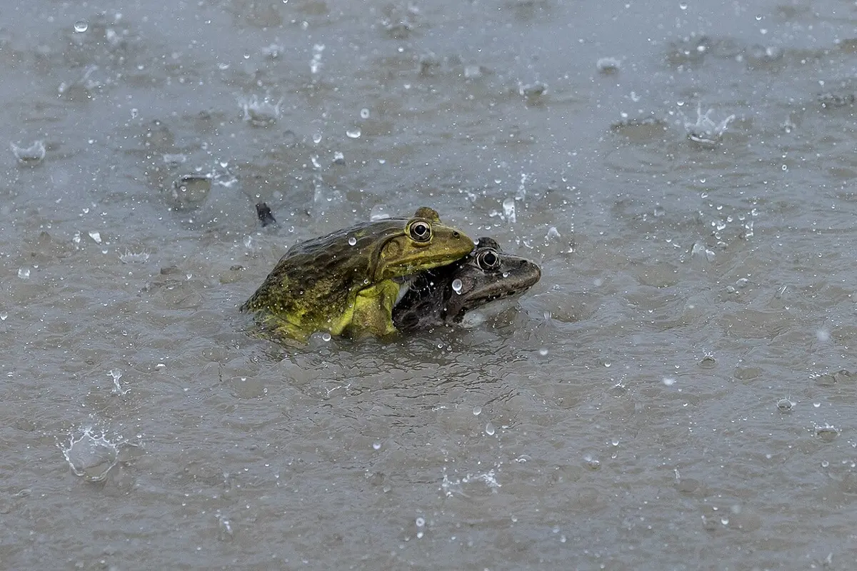
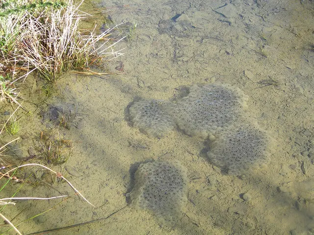
In the bullfrog, where does fertilization happen?
Pick an answer to see why.
Exercise: Reproductive system
A. Tick the correct alternative.
- Which part of a sperm gives it the energy to move?
Pick an answer to see why.
- Fertilization begins when a sperm gets through the egg’s coverings. How does it do so?
Pick an answer to see why.
- Bidder’s canal is in:
Pick an answer to see why.
- The making of an egg is called:
Pick an answer to see why.
- A female frog’s belly bulges in the breeding season because:
Pick an answer to see why.
- A frog’s egg is mesolecithal because of the … yolk in it.
Pick an answer to see why.
- The outer covering of a frog’s unfertilized egg just after ovulation is:
Pick an answer to see why.
- A frog’s egg is telolecithal. This means:
Pick an answer to see why.
- Testosterone brings on a male frog’s breeding features and controls much else. Which cells of the testis mainly make it?
Pick an answer to see why.
B. Answer the following questions.
What is sexual dimorphism? How is a male frog told apart from a female?
Answer
Draw a neat, labeled diagram of the male reproductive system of a frog and explain it.
Answer
Briefly describe the structure of a frog’s sperm, with a diagram.
Answer
Draw a neat, labeled diagram of a T.S. of a frog’s testis.
Answer
Why is the male frog’s ureter called a urinogenital duct, unlike the female’s?
Answer
Sperm: S. J. Holmes, The Biology of the Frog (1906), public domain
Look at Figure 26.28.
- i.Name the cell and label the parts A, B, C and D.
- ii.What do A and D do?
- iii.Name the organelle in C that helps the egg divide (cleave) after fertilization.
Answer
List the female reproductive organs of a frog and describe them, with a well-labeled diagram.
Answer
Briefly describe the structure of a frog’s unfertilized egg.
Answer
Printable PDFs
Quiz yourself: Classification of Living Beings, Grades 5–12 →
Practice every question in the book, and keep track of the ones you miss →
A companion to your textbook, not a replacement for it: study from the book your school uses, and where the two differ, follow your teacher. Found a mistake? Please tell us.