
In 1881, a year before he died, Charles Darwin published his last book. It was not about apes or finches but about earthworms, which he had watched for some forty years. He set chalk on a field and dug it up decades later to find it buried under fresh soil the worms had brought up; he played a bassoon and a piano to them (they took no notice of the sound, but felt the piano’s vibrations through a pot of soil). He worked out that the worms of an English field pass something like ten tonnes of earth per acre through their bodies every year. “It may be doubted,” he wrote, “whether there are many other animals which have played so important a part in the history of the world.”
This unit looks closely at one earthworm, the common Indian earthworm, Pheretima posthuma, from its skin to its nerves. Almost every one of its organs sits in a fixed segment, so learning the earthworm is partly learning a map: which organ, in which ring.
Observe and discuss
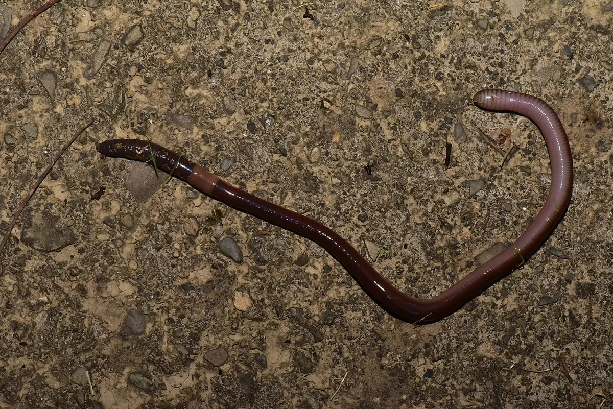
8.3.1 General introduction
Systematic position
| Rank | Name |
|---|---|
| Kingdom | Animalia |
| Phylum | Annelida |
| Class | Oligochaeta |
| Genus | Pheretima |
| Species | posthuma |
| Common name | Earthworm |
The earthworm belongs to the phylum Annelida, the segmented worms, and to the class Oligochaeta (Greek oligos, few; chaete, bristle): annelids with a few bristles on each segment, a clitellum, and no head or parapodia. Its scientific name is Pheretima posthuma.
Earthworms live in moist soil, in burrows. They have no special breathing organs, no lungs or gills: they take in oxygen and give out carbon dioxide through their moist skin. Breathing through the skin is called cutaneous respiration. Glands in the skin pour out mucus, which keeps it moist.
The earthworm is free-living and omnivorous: it swallows soil rich in decaying organic matter, and digests dead leaves, insect larvae, protozoa, the remains of arthropods’ exoskeletons and the microbes that live on all of them. A short way behind its front end is a thick, ring-like band of glands, the clitellum, which makes the cocoon. By burrowing and by its droppings it makes the soil richer, which is why it is called the farmer’s friend.
8.3.2 Habit and habitat
Earthworms are terrestrial. They live in burrows in moist soil rich in decaying organic matter, mostly in irrigated farmland, gardens and lawns, and avoid dry, sandy or very acid soil.
They are nocturnal: by day they stay in their burrows, and at night they come out to look for food, to find new places to live and to mate. After heavy rain you see them in large numbers on the surface. The rainy season is their breeding season, and the rain fills their burrows with water, so they come up.
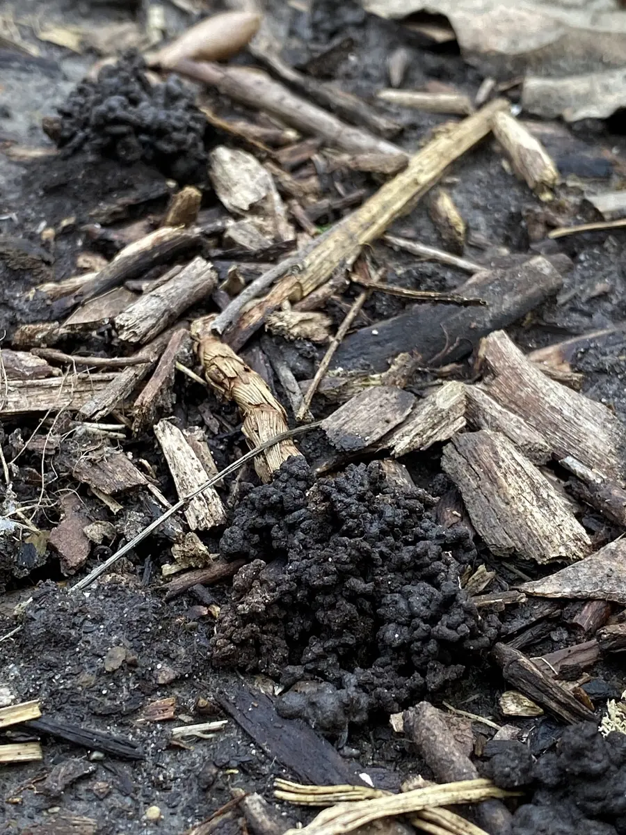
An earthworm digs its burrow by eating its way through the soil. It pushes its pointed front end into a crack, swallows the soil in front of it and moves down bit by bit. Its food is the decaying organic matter in the soil it swallows; what it can’t digest, mostly soil, it passes out as small heaps of pellets, the worm castings, often seen at the mouth of a burrow.
Earthworms are hermaphrodite: each worm has both male and female organs. Yet a worm does not fertilize itself. Two mature worms always mate (copulate) and exchange sperm. Fertilization is external: it happens outside the body, inside the cocoon. Development is direct: no larva; a tiny worm comes out of the cocoon.
Why do earthworms come up on the surface in large numbers after heavy rain?
Pick an answer to see why.
8.3.3 External features (morphology)
1. Shape, size and color
The body of Pheretima is vermiform (worm-shaped): long, narrow and cylindrical, and bilaterally symmetrical. It is pointed at the front (anterior) end and blunter at the back (posterior). A mature worm is about 15 cm long and 3–5 mm thick.
It is a shiny dark brown, from the pigment porphyrin in its body wall, which also shields it from the harmful ultraviolet rays of the Sun. The dorsal (upper) surface is darker than the ventral (lower) one, and along the middle of the back runs a dark line: the dorsal blood vessel, seen through the skin.
2. Segmentation
The soft body is divided into about 100–120 similar rings, the segments (somites or metameres). On the outside they are separated by grooves, the intersegmental grooves; inside, by thin partitions, the septa (singular septum). Because the outer rings match the inner divisions, and each segment repeats the same set of organs, the segmentation is true or metameric (metamerism).
3. Prostomium and peristomium
The earthworm has no distinct head and no eyes or tentacles. Its first segment holds the mouth and is called the peristomium (peri, around; stoma, mouth). Above the mouth, the peristomium carries a fleshy lobe that sticks forward over it, the prostomium (pro, in front of). The prostomium is not counted as a segment. It is the worm’s boring organ, which pushes into cracks in the soil, and it is sensitive to touch.
4. Clitellum
In a mature worm a thick, smooth band of glands surrounds segments 14, 15 and 16: the clitellum or cingulum. It has three kinds of gland cells: mucus-secreting, albumen-secreting and cocoon-forming cells. Being plain and swollen, it has no setae and no visible grooves between its three segments.
The clitellum divides the body into three regions:
- pre-clitellar region: segments 1 to 13;
- clitellar region: segments 14 to 16;
- post-clitellar region: segment 17 to the last.
5. Setae
Every segment except the first, the last and the three of the clitellum carries a ring of small bristles, the setae (singular seta), made of chitin and pointing backwards. They sit in a single row round the middle of each segment, about 80–120 in each ring. Bristles in a complete ring round each segment are called the perichaetine arrangement (peri, around; chaete, bristle).
A seta is pale yellow and shaped like a long, slim letter ‘S’, swollen near its middle (the nodulus). Each sits in a pocket of skin, the setal sac, and is moved by two small muscles: a protractor muscle pushes it out and a retractor muscle pulls it in. The setae grip the soil when the worm moves, so they are its locomotory organs.
How an earthworm crawls
The setae are only half the story. Under the skin lie two layers of muscle at right angles: circular muscles, which run round the body, and longitudinal muscles, which run along it. Each segment is a closed bag of coelomic fluid, and a liquid can’t be squeezed smaller.
- When a segment’s circular muscles contract, it gets thinner; its fluid has to go somewhere, so the segment gets longer. Its setae are pulled in and it slides forward.
- When its longitudinal muscles contract, it gets shorter and fatter, and its setae stick out and grip the burrow’s wall, holding that part still.
Waves of these two contractions run from the front of the worm to the back. The fat, anchored segments hold on while the thin ones in front reach forward, so the worm moves ahead a little with each wave. The fluid in the segments, pushed by muscles, works as a hydrostatic skeleton (hydroskeleton).
When the circular muscles of a segment of an earthworm contract, the segment becomes:
Pick an answer to see why.
6. External apertures
The body wall has these openings, as in Figure 25.5 and Figure 25.6:
- i.Mouth: a crescent-shaped opening, the front-most one, on the ventral side of the first segment, the peristomium, under the prostomium.
- ii.Spermathecal pores: four pairs of small openings of the spermathecae (sperm-storing sacs), one pair in each of the intersegmental grooves 5/6, 6/7, 7/8 and 8/9, ventro-laterally (low on each side).
- iii.Nephridiopores: very many tiny openings of the integumentary nephridia, scattered all over the body except the first two segments. Through them the worm passes out nitrogen-containing wastes taken from its blood.
- iv.Dorsal pores: tiny openings along the mid-dorsal line, one in each intersegmental groove from 12/13 to the end, except the last groove. They open from the coelom, so coelomic fluid oozes out through them and keeps the skin moist.
- v.Female genital aperture: a single opening in the middle of the ventral side of segment 14, the end of the common oviduct, through which the eggs (ova) are laid.
- vi.Male genital apertures: a pair of crescent-shaped openings on the ventro-lateral sides of segment 18, through which the sperm pass out at mating.
- vii.Anus: a vertical slit at the end of the last segment, the anal segment or pygidium, through which the feces (castings) leave the gut.
7. Genital papillae
There are two pairs of genital papillae, rounded swellings on the ventro-lateral surface, one pair on segment 17 and one on segment 19, just in front of and behind the male apertures. The accessory glands open on them, and they help two worms hold on to each other during mating.
In which segment of Pheretima is the single female genital aperture?
Pick an answer to see why.
Coelomic fluid oozes out on the earthworm’s back through the:
Pick an answer to see why.
Body wall of the earthworm
The body wall is thin, soft, elastic and moist. From outside in, it has four layers: the cuticle, the epidermis, the muscle layer and the parietal coelomic epithelium.
1. Cuticle. A thin, elastic, non-living, finely striated outer covering. It is made of two layers of collagen fibers in a matrix of polysaccharide with a little gelatin, and is secreted by the epidermis beneath it. It is pierced by many fine pores, through which the epidermal glands and the nephridia open.
2. Epidermis. A single layer of cells under the cuticle, resting on a basement membrane. It has four kinds of cells:
- Supporting cells: tall columns that make up most of the epidermis and secrete the cuticle.
- Gland cells: mucous cells, which make mucus, and albumen cells, which make albumen. Their secretions keep the skin moist and slippery and help to cement the walls of the burrow.
- Basal cells: small, cone-shaped cells at the bottom of the layer, between the others. They replace worn-out supporting and gland cells, so they are also called replacing cells.
- Sensory cells: in groups, with fine hair-like ends sticking out through the cuticle. They sense light, chemicals and touch (photoreceptors, chemoreceptors and touch receptors).
3. Muscle layer. Under the epidermis:
- an outer, thin layer of circular muscle fibers, running round the body, with scattered granules of porphyrin, which gives the skin its brown color;
- an inner, thick layer of longitudinal muscle fibers, running along the body, in parallel bundles separated by connective tissue and strengthened by collagen;
- under the longitudinal layer, another thin strip of circular muscle.
The circular and longitudinal muscles work against each other (they are antagonistic): when one contracts, the other relaxes, as in the crawling widget above.
4. Parietal coelomic epithelium. A single layer of thin, flat (squamous) cells, which forms the outer lining of the coelom.
Coelom in the earthworm
The earthworm has a true coelom, the fluid-filled space between the gut and the body wall, lined on both sides by mesoderm. It forms in the embryo by the splitting of the mesoderm, so it is a schizocoel (Unit 24, Body cavity).
The coelom is divided into coelomic chambers, one to a segment, by the muscular septa between the segments:
- there are no septa in the first four segments; the first septum, 4/5, is thin;
- between segments 8 and 10, round the gizzard, there is only one septum instead of two;
- the septa from 4/5 to 13/14 are without holes;
- the septa from 14/15 to the last have holes with sphincters (rings of muscle that open and close them), so coelomic fluid can pass from one chamber to the next.
The coelom is filled with coelomic fluid: milky white or colorless and alkaline, made of water, salts, proteins and floating cells, the coelomic corpuscles. They are of four kinds:
- i.Phagocytes (amoebocytes, granulocytes): the largest and most numerous, nucleated and amoeba-like. They engulf bacteria, which is why they are called phagocytes (“eating cells”).
- ii.Mucocytes: long cells, a broad fan at one end and a narrow nucleated part at the other.
- iii.Circular nucleated cells: fewer, round, nucleated, with clear cytoplasm.
- iv.Chloragogen cells (“yellow cells”): small, star-shaped cells, full of yellow granules, that grow on the outside of the intestine and break off into the coelom. They take wastes out of the coelomic fluid and turn ammonia into urea, and they store food as glycogen and fat, so they work much like the liver of a vertebrate.
Functions of the coelomic fluid
- i.It works as a hydrostatic skeleton: it keeps the body firm and stiff, which the muscles push against when the worm crawls and burrows.
- ii.It oozes out through the dorsal pores and keeps the skin moist for cutaneous respiration.
- iii.Its phagocytes kill bacteria, so it protects the body and skin against infection.
Which coelomic corpuscles of the earthworm store glycogen and are compared to the liver of vertebrates?
Pick an answer to see why.
8.3.4 Digestive system
The digestive system turns complex food into simple forms that can be absorbed, takes them into the body, and gets rid of what is left as feces. It has two parts: the alimentary canal and the digestive glands.
The earthworm’s alimentary canal is a long, complete, straight tube of varying width, from the mouth to the anus; it is also called the gastrointestinal tract. It has these parts, each in its own segments:
1. Mouth and buccal chamber (segments 1–3). The crescent-shaped mouth, under the prostomium, opens into a short, narrow, thin-walled buccal chamber (buccal cavity), which reaches to the middle of segment 3. It can be pushed out (everted) through the mouth to grip food. Its inner lining is folded lengthwise.
2. Pharynx (segments 3–4). A wide, pear-shaped, muscular chamber behind the buccal chamber, separated from it by a groove. It runs from the middle of segment 3 to segment 4.
Its roof is thick and forms the pharyngeal bulb, which holds the salivary glands, made of deeply staining (chromophil) cells. Two horizontal shelves from its side walls divide its cavity, incompletely, into an upper salivary chamber, into which the glands pour saliva, and a lower ventral conducting chamber, through which the food simply passes. Saliva has mucus, which wets and lubricates the food, and proteolytic (protein-digesting) enzymes.
The pharynx is tied to the body wall by muscle strands. As they contract and relax, they widen and narrow the pharynx, so it sucks in soil and food and pumps it backwards.
Body outline: traced from the British Museum (public domain)
3. Esophagus (segments 5–7). A short, narrow, thin-walled tube without glands. Food is pushed through it by peristalsis, waves of muscle contraction.
4. Gizzard (segment 8, or 8–9). A distinct, oval, hard, thick-walled organ, its wall mostly circular muscle, its cavity lined with cuticle. It grinds the food, with the grains of sand swallowed with it, into a fine paste: it is the grinding organ.
Body outline: traced from the British Museum (public domain)
5. Stomach (segments 9–14). A short, narrow, thin-walled tube with a sphincter at each end. Its lining is glandular and rich in blood vessels, and folded across. In its wall are calciferous glands, which take excess calcium and carbon dioxide out of the blood and pass them into the gut as solid calcium carbonate (calcite). This keeps the calcium in the blood steady, and the calcium carbonate neutralizes the humic acids of the decaying leaves the worm eats.
6. Intestine (segment 15 to the last). A long, wide, thin-walled tube that runs to the end of the body. It looks beaded, because it is pinched in where each septum crosses it. It has three regions:
- i.Pre-typhlosolar region (segments 15–26). Its lining is glandular, rich in blood vessels and folded into tiny projections, the villi. In segment 26 a pair of short, cone-shaped intestinal ceca (singular cecum) grow out from its sides and point forwards, reaching to about segment 23. They are rich in blood vessels, have villi and secrete the enzyme amylase.
- ii.Typhlosolar region (segment 27 to all but the last 23–25). Along the middle of its roof hangs a long fold, the typhlosole, glandular and full of blood vessels. It is like one huge villus: it greatly increases the inner surface of the intestine, and most digestion and absorption happens here.
- iii.Post-typhlosolar region (the last 23–25 segments). Also called the rectum. Thin-walled and rich in blood vessels; it has no typhlosole, villi or gland cells, only lengthwise folds. It holds small pellets of mud, the feces.
7. Anus. The intestine opens to the outside through the anus on the last segment. The worm castings are passed out through it.
Body outline: traced from the British Museum (public domain)
Physiology of digestion
Digestion turns complex food into simple forms that can be absorbed. It is mechanical (breaking large pieces into small ones, so enzymes can reach more of it) and chemical (enzymes breaking carbohydrates, proteins and fats down to glucose and other sugars, amino acids, fatty acids and glycerol).
1. Food. The earthworm is omnivorous and detritivorous. It swallows soil rich in decaying organic matter such as rotting leaves, along with nematodes, protozoa, insects’ eggs and larvae and other microbes; it can also eat leaves, grass and algae directly.
2. Feeding. The buccal chamber is pushed out through the mouth, grips soil and food, and is pulled back; the sucking and pumping of the muscular pharynx then draws the soil and food in.
3. Digestion. Nothing is digested in the buccal chamber.
- In the pharynx, saliva is added: its mucus lubricates the food, and its proteolytic enzymes start breaking proteins into proteoses and peptones.
- The food passes down the esophagus by peristalsis into the gizzard, whose muscular wall and cuticle-lined cavity grind it into a fine paste.
- In the stomach, the digestion of proteins continues.
- In the intestine, enzymes from the intestine and the ceca finish the work:
| Enzyme | Acts on | Gives |
|---|---|---|
| Amylase | starch (carbohydrates) | simple sugars |
| Protease | proteins, proteoses and peptones | amino acids |
| Lipase | fats | fatty acids and glycerol |
| Chitinase | chitin, from the exoskeletons of arthropods | sugars |
| Cellulase | cellulose, from plant cell walls | glucose |
4. Absorption and egestion. The digested food is absorbed through the villi of the intestine, above all through the typhlosole, into the blood capillaries in the intestinal wall, and carried round the body. So the intestine is where digestion is finished and food absorbed. What can’t be digested, with the soil, is passed out through the anus as small round pellets of mud, the worm castings.
In the earthworm, most of the digested food is absorbed in the:
Pick an answer to see why.
What is the main use of the gizzard to the earthworm?
Pick an answer to see why.
8.3.5 Excretory (nephridial) system
Every living thing makes wastes in its body’s chemistry, and these must be removed to keep the inside of the body steady and healthy. Excretion is the removal of these metabolic wastes, especially the nitrogen-containing ones. The earthworm is both ammonotelic (it gets rid of ammonia) and ureotelic (it gets rid of urea).
It excretes by nephridia (singular nephridium): microscopic, long, thin, coiled tubes, arranged segment by segment. In Pheretima they are tiny and very many, so they are called micronephridia, unlike the large meganephridia of Nereis (one pair in each segment) and of the leech.
By where they are, the nephridia of Pheretima are of three kinds:
- Pharyngeal nephridia: in segments 4, 5 and 6.
- Integumentary nephridia: on the body wall of every segment except the first two.
- Septal nephridia: on both faces of each septum from 15/16 to the last.
1. Pharyngeal nephridia
These lie as three pairs of tufts, one pair on each side of the pharynx and esophagus in segments 4, 5 and 6. Each tuft has hundreds of fine, coiled, branched tubules, with blood glands among them. The tubules of a tuft join into one thick-walled common duct, which runs forward beside the ventral nerve cord. The ducts from segments 4 and 5 open into the pharynx, and those from segment 6 into the buccal chamber in segment 2.
So they pour their wastes into the gut: they are enteronephric (or endonephric) nephridia. They have no nephrostome (no funnel open to the coelom), so they are closed: they take their wastes only from the blood in the capillaries round them.
2. Integumentary nephridia
These are the smallest: microscopic, V-shaped tubules, attached to the inside of the body wall of every segment except the first two. There are about 200–250 in a segment, but in each segment of the clitellum about 2,000–2,500, so the clitellum is called the forest of nephridia.
They have no nephrostome (they are closed), and they take their wastes only from the blood. Each opens to the outside by its own tiny nephridiopore on the skin, so they pour their wastes outside: they are exonephric (ectonephric) nephridia.
3. Septal nephridia
These are the largest and the most typical or advanced nephridia of the earthworm, because they alone have a nephrostome, a funnel open to the coelom. They hang freely in the coelom on both faces of each septum from 15/16 backwards, about 40–50 on each face, so 80–100 on each septum; the first septum, 15/16, carries only about 40–50. Like the pharyngeal ones, they pour their wastes into the gut: they are enteronephric.
Each septal nephridium has three parts:
- i.Nephrostome (nephridiostome). The open, ciliated funnel at its front end, which faces the coelom of the segment in front. Its oval mouth has an upper lip of about eight ciliated marginal cells round a large central cell, and a lower lip of about four ciliated cells. The cilia beat all the time and draw coelomic fluid, with its wastes, into the tube.
- ii.Body. A short, thin, ciliated neck joins the nephrostome to the body, which has two parts: a short straight lobe and a long twisted loop. The loop is made of a proximal limb (joined to the neck’s end of the tube) and a distal limb, wound round each other in a spiral of 9–13 twists, with a narrow apical part at its tip.
- iii.Terminal duct. A short, narrow tube from the distal limb, which opens into the septal excretory canal.
Inside the gland cells of the nephridium runs the nephridial tube, which passes up and down the body several times. In cross-section the body shows several channels side by side: four in the straight lobe, three each in the proximal and distal limbs, two in the apical part, and one in the neck and in the terminal duct.
Traced from K. N. Bahl, 1919 (public domain)
Where the septal nephridia empty. Two supra-intestinal excretory ducts run along the top of the intestine from segment 15 to the end. On each septum, a septal excretory canal on each side collects the terminal ducts of all the septal nephridia of that side and leads up into the supra-intestinal duct of its side. In each segment the supra-intestinal ducts open into the intestine by fine ductules, each with a sphincter. So:
septal nephridia → terminal ducts → septal excretory canals → supra-intestinal excretory ducts → intestine → out with the feces through the anus.
Physiology of excretion
The fluid an earthworm excretes carries about 40% ammonia, 50% urea and 0.6% amino acids, with other compounds in small amounts. A well-fed worm passes out more ammonia; a starving one more urea.
- The gland cells of the pharyngeal and integumentary nephridia take nitrogen-containing wastes and water out of the blood. The integumentary nephridia pass them straight out through the nephridiopores; the pharyngeal ones pass them into the gut, and they leave through the anus.
- The septal nephridia take wastes from the coelomic fluid too: the cilia of their nephrostomes keep drawing the fluid in. Their wastes go into the gut and leave with the feces.
Which nephridia of the earthworm take wastes from the coelomic fluid?
Pick an answer to see why.
8.3.6 Nervous system
The nervous system controls and co-ordinates the body: it picks up stimuli, makes the body respond, and keeps its many activities working together. In Pheretima it is well developed and concentrated. It has three parts: the central, the peripheral and the sympathetic nervous system.
1. Central nervous system (CNS)
The CNS is a nerve ring at the front, and the ventral nerve cord that runs back from it.
a. Nerve ring. It surrounds the pharynx in segments 3 and 4, and is made of:
- the supra-pharyngeal (cerebral) ganglia: a pair of white, pear-shaped ganglia fused together, the worm’s brain, lying on the dorsal side in the groove between the buccal chamber and the pharynx, in segment 3;
- a pair of thick circum-pharyngeal (peri-pharyngeal) connectives, which run down from the sides of the brain, one round each side of the pharynx;
- the sub-pharyngeal ganglia, a fused pair under the pharynx in segment 4, where the two connectives meet on the ventral side.
After a drawing by Looie496 (public domain)
b. Ventral nerve cord. A long, solid rod that runs back from the sub-pharyngeal ganglia along the mid-ventral line, under the gut, to the last segment. In each segment it swells into a segmental ganglion.
It looks single, but it is double: two cords, right and left, side by side and fused together, and each segmental ganglion is a fused pair of ganglia, one from each. Inside are nerve cells and nerve fibers, among them three giant fibers along its top, which carry signals very fast. It is wrapped in a sheath of three layers: an outer visceral peritoneum, a middle layer of longitudinal muscle fibers, and an inner, thick, fibrous epineurium.
2. Peripheral nervous system
The nerves that leave the CNS make up the peripheral nervous system:
- from each cerebral ganglion, about 8–10 nerves to the prostomium and the buccal chamber;
- from the circum-pharyngeal connectives, two pairs of nerves to the first segment and the buccal chamber;
- from the sub-pharyngeal ganglia, three pairs of nerves to segments 2, 3 and 4;
- from each segmental ganglion, three pairs of segmental nerves: one pair in front of the ganglion and two pairs behind it. They supply the body wall, the gut wall and the other organs of that segment.
All these nerves are mixed: each carries signals both to the CNS (afferent) and from it (efferent).
3. Sympathetic nervous system
A fine network (plexus) of nerves under the epidermis and in the muscles of the body wall, and on the wall of the gut and other organs. These plexuses are joined to the circum-pharyngeal connectives.
Sense organs
The earthworm’s sense organs are simple, single cells or small groups of cells of the epidermis (from the ectoderm), but there are many of them:
- i.Epidermal receptors: all over the epidermis, most on the sides and the ventral surface. Each cell has fine hair-like ends at the surface and a nerve fiber at its base. They respond to touch and chemicals.
- ii.Buccal receptors: in the buccal chamber; they taste and smell the food.
- iii.Photoreceptors: on the dorsal surface, most on the prostomium and peristomium, fewer towards the back, none on the clitellum. Each is a cell with a clear lens-like body in it. They tell how bright the light is, so the worm can stay out of it.
Where is the brain of the earthworm?
Pick an answer to see why.
8.3.7 Reproductive system
Reproduction is how living things produce young of their own kind, so that the species goes on. The earthworm is hermaphrodite (monoecious): one worm has both male and female organs. Yet it can’t fertilize itself, because it is protandrous: its male organs ripen first, before its own eggs are ready. So two mature worms mate (copulate) and exchange sperm (cross-fertilization), and cocoons are made afterwards.
A. Male reproductive organs
1. Testes. Two pairs of tiny, white, lobed testes, one pair in segment 10 and one in segment 11, each lying low (ventro-laterally) beside the ventral nerve cord and attached to the front wall of its testis sac. Each has a narrow base with 4–8 finger-like lobes holding round balls of sperm mother cells (spermatogonia). These break off into the testis sac and pass into the seminal vesicles to mature into sperm (spermatozoa).
2. Testis sacs. Two wide, white, thin-walled sacs filled with fluid, in segments 10 and 11, below the gut, which enclose the testes. Each also encloses a pair of spermiducal funnels. The sac of segment 11 is the larger. Each sac opens by narrow ducts into a pair of seminal vesicles.
3. Seminal vesicles. Two pairs of large, white sacs, one pair in segment 11 and one in segment 12; those of 12 are the larger and are not enclosed in a testis sac. They grow out from the septa, so they are also called septal pouches. The seminal vesicles of segment 11 receive from the testis sac of segment 10, and those of 12 from the testis sac of 11. In them the sperm mother cells mature into sperm.
4. Spermiducal funnels. Two pairs of ciliated cups with folded rims, one under each testis in segments 10 and 11, inside the testis sacs. Mature sperm move back from the seminal vesicles into the testis sacs and enter the vasa deferentia through these funnels.
5. Vasa deferentia. Each funnel leads into a fine, thread-like, ciliated sperm duct (vas deferens; plural vasa deferentia). The two of each side run back close together beside the nerve cord, along the ventral body wall, to segment 18, where they join the prostatic duct.
6. Prostate glands. A pair of large, dull white, flat, lobed glands on either side of the gut, from about segment 16 to 21. Each has a large glandular part of many lobules and a small non-glandular part of fine ductules, which join into a prostatic duct. In segment 18, the prostatic duct and the two vasa deferentia of the same side run inside one muscular sheath, as a single tube, the common prostatic and spermatic duct. It curves down and opens by the male genital aperture on segment 18. The prostatic fluid is passed out with the sperm; its exact role is not well understood.
7. Accessory glands. Two pairs of round, white glands, one pair in segment 17 and one in 19, one on each side of the nerve cord. They open by fine ducts on the genital papillae, and their sticky secretion helps the two worms hold together at mating.
B. Female reproductive organs
1. Ovaries. One pair of small, white, lobed ovaries in segment 13, hanging from the septum 12/13, one on each side of the ventral nerve cord. Each has finger-like lobes in which eggs (ova) at every stage lie in a row: the youngest near the base, the largest and ripest at the free tips.
2. Oviducal funnels. A pair of large, saucer-shaped funnels with folded, ciliated rims, one under each ovary in segment 13.
3. Oviducts. Each funnel leads into a short, cone-shaped, ciliated oviduct. The two run back through the septum 13/14 and join into a common oviduct, which opens by the single female genital aperture in the middle of the ventral side of segment 14.
4. Spermathecae. Four pairs of small, flask-shaped sacs, one pair in each of segments 6, 7, 8 and 9, ventro-laterally. Each has a broad, pear-shaped ampulla and a short, narrow neck, with a small blind pouch, the diverticulum, on its side. At mating the sperm received from the partner are stored in the diverticula; the ampulla feeds them and keeps them alive until the worm’s own eggs are ripe. Each pair opens to the outside by the spermathecal pores in the grooves 5/6, 6/7, 7/8 and 8/9.
Spermatheca: after a photograph by Nguyen, Lam, Trinh and Nguyen, 2020, ZooKeys, CC BY 4.0
In Pheretima, sperm from the partner are stored in the:
Pick an answer to see why.
An earthworm is hermaphrodite, yet it does not fertilize itself, because it is:
Pick an answer to see why.
8.3.8 Copulation and cocoon formation
Copulation
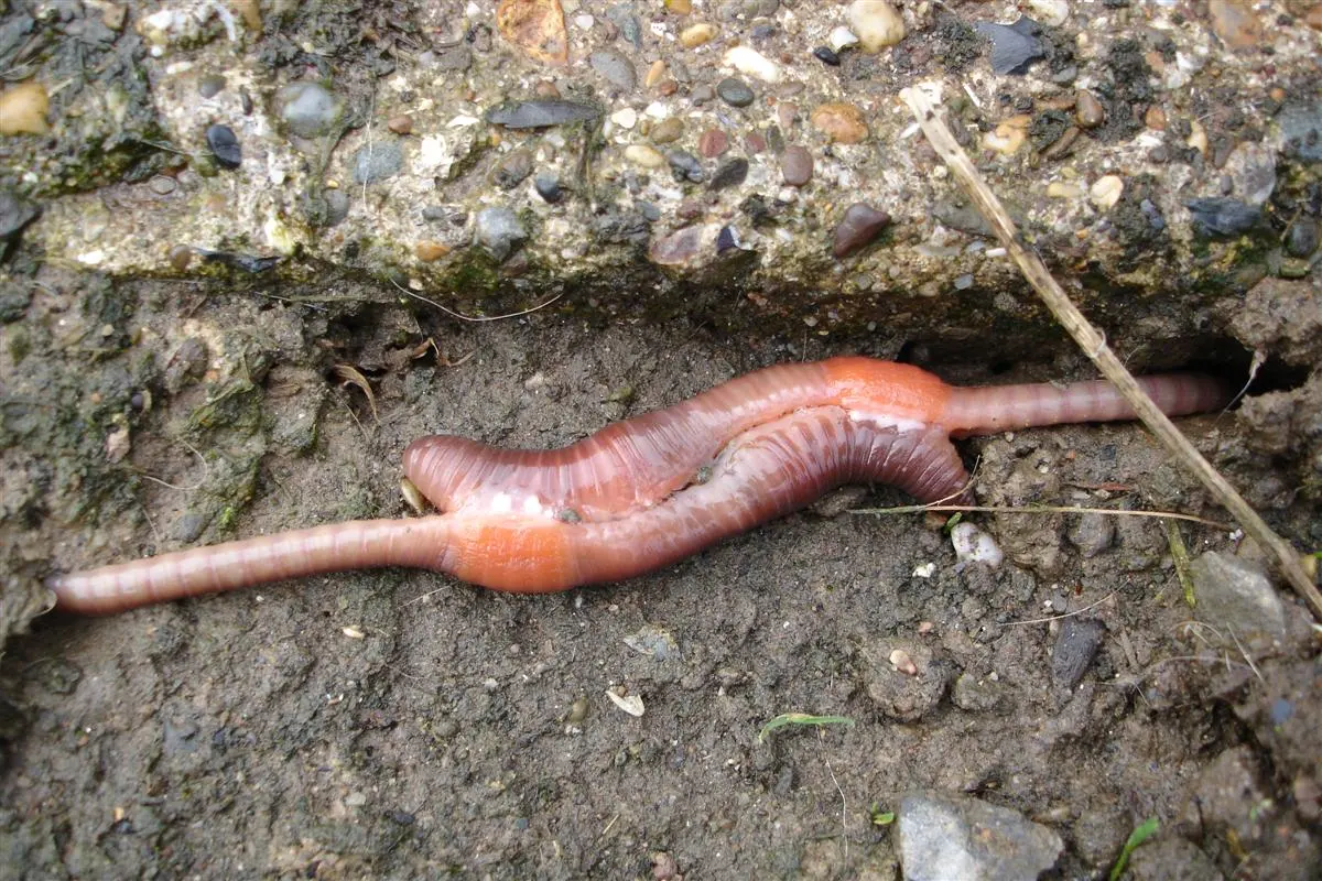
Earthworms mate in the rainy season, from about July to October, at night, for about an hour. Two mature worms lie with their ventral surfaces together, their heads pointing opposite ways. They lie so that the male apertures (segment 18) of each worm are against the spermathecal pores (segments 6 to 9) of the other, as in Figure 25.20. The skin round each male aperture rises into a little papilla, which acts as a copulatory organ and is pushed into the other worm’s spermathecal pores. In this way each worm passes sperm and prostatic fluid to the other, and the two then separate. The sperm received are kept alive in the spermathecae until the worm’s own eggs are ripe.
Cocoon formation
Some time after mating, when the worm’s ovaries are ripe, it makes cocoons. The clitellum’s three kinds of gland cells each do a job:
- The cocoon-forming glands of the clitellum pour out a sticky substance, which hardens in the air into a membranous band (girdle) round the clitellum. The albumen glands fill it with albumen, a food store for the young.
- The worm backs out of the band, so the band slides forwards over its body.
- As the band passes segment 14, several eggs (ova), still unfertilized, are laid into it through the female aperture.
- As it passes segments 9 to 6, sperm from the spermathecae are poured into it through the spermathecal pores.
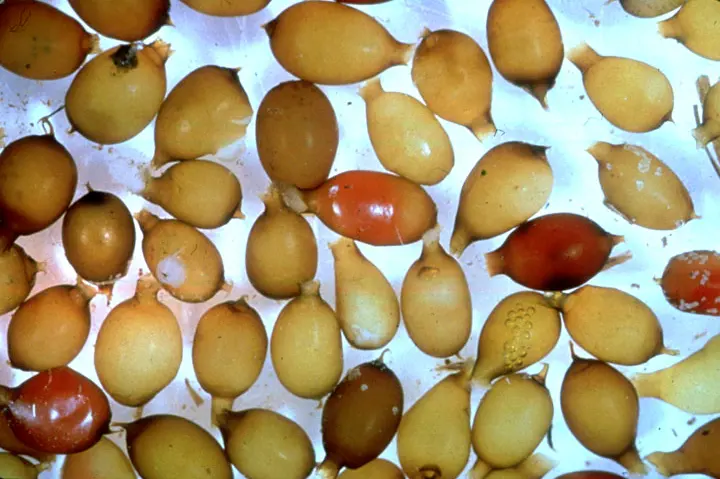
- The band slips off the front end, and its elastic ends close up, making a small, light yellow, oval cocoon. Many cocoons are made, one after another, after each mating.
The eggs are fertilized inside the cocoon, after it is laid in the soil: fertilization is external (outside the body). Of the many eggs, usually only one grows into a young worm. The zygote divides by holoblastic, unequal cleavage, and development is direct: there is no larva. After about 15–20 days a tiny worm comes out of the cocoon.
During cocoon formation, the eggs enter the cocoon band:
Pick an answer to see why.
8.3.9 Economic importance of the earthworm
Useful
- Food for other animals: birds, lizards, frogs, snakes and fish eat earthworms, and anglers use them as bait.
- Better soil: burrowing loosens the soil and lets air and water in, and the castings mix organic matter and minerals into it. Its physical and chemical properties improve, which helps farmers.
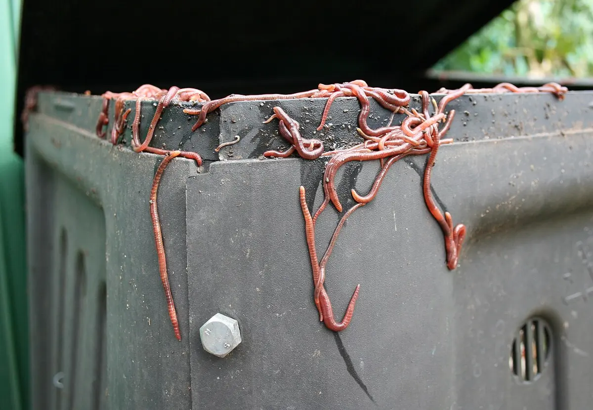
- Vermicomposting: earthworms are used to turn organic waste into compost manure, vermicompost.
- Traditional medicine: in the past, earthworm preparations were used for piles, jaundice, rheumatic fever, gallstones and infertility. These are traditional uses; there is no good evidence that they cure these diseases.
- Study: as a common, easily dissected invertebrate, the earthworm is the type animal for learning the anatomy and physiology of annelids.
Harmful
- Many burrows in irrigated fields can let the water seep away, which can lower the crop yield.
- Soil loosened by earthworms is washed away more easily from sloping land.
- Earthworms coming up to the surface can carry parasites of other animals: the poultry tapeworm Amoebotaenia, which grows its larvae in earthworms, the gapeworm of poultry and other nematodes; and bacteria and viruses in their bodies can infect animals that eat them.
- Some species damage the roots of crops such as rice (paddy) and betel vine.
Which earthworm is most used for vermicomposting?
Pick an answer to see why.
Exercise
A. Tick the correct alternative.
- Which pigment gives the earthworm’s body its glistening brown color?
Pick an answer to see why.
- In which of the following structures is the mouth located?
Pick an answer to see why.
- Which kind of segmentation is found in the body of the earthworm?
Pick an answer to see why.
- When two mature earthworms mate, they exchange sperm. Which surfaces of their bodies come together?
Pick an answer to see why.
- Which part of the earthworm holds the fertilized eggs?
Pick an answer to see why.
- In which segments are the seminal vesicles?
Pick an answer to see why.
- How does the earthworm’s excretory system help to make the soil fertile?
Pick an answer to see why.
- Which part mainly crushes the food in the earthworm’s digestive system?
Pick an answer to see why.
- In which segments is the nerve ring?
Pick an answer to see why.
- What is the feeding habit of the earthworm?
Pick an answer to see why.
- What are the earthworm’s organs of locomotion?
Pick an answer to see why.
- Which of these are exonephric nephridia?
Pick an answer to see why.
- Which statement best describes absorption in the earthworm’s alimentary canal?
Pick an answer to see why.
- Which part of the earthworm is called the forest of nephridia?
Pick an answer to see why.
- Which cells are called yellow cells?
Pick an answer to see why.
- Cocoon formation is a special feature of the earthworm’s life. Which of these is true of the cocoon?
Pick an answer to see why.
- Where are the dorsal pores?
Pick an answer to see why.
- Which nephridia take wastes from the coelomic fluid?
Pick an answer to see why.
- Earthworm nephridia are with or without a nephrostome. Which have a nephrostome?
Pick an answer to see why.
B. Answer the following questions.
Write a short note on the habit and habitat of Pheretima posthuma.
Answer
What is morphology? Write the morphological features of the earthworm.
Answer
Draw a neat, labeled diagram of the T.S. of the body wall of the earthworm through the setae.
Answer
The earthworm is omnivorous and detritivorous. Describe the process of digestion of food in its digestive tract.
Answer
Write a short note on the central nervous system of the earthworm.
Answer
How do you differentiate septal nephridia from pharyngeal nephridia?
Answer
Look at the diagram above and answer these questions.
- i.Name the parts A, B, C, D and E.
- ii.In which part do the sperm mature?
- iii.What are the functions of parts C and E?
Answer
Draw a neat, labeled diagram of the T.S. of the earthworm through the pharynx.
Answer
Write four differences between the male and the female reproductive systems of the earthworm.
Answer
What is the typhlosolar region of the intestine of the earthworm? How is it different from the post-typhlosolar region?
Answer
Saumyata said that the earthworm is a hermaphrodite animal, which fertilizes itself and has no need to mate. Is she right? Give your opinion.
Answer
How do copulation and cocoon formation take place in the earthworm? Describe the process.
Answer
Body outline: traced from the British Museum (public domain)
Draw a well-labeled diagram of the T.S. of the earthworm through the gizzard.
Answer
What is the economic importance of the earthworm?
Answer
After a drawing by Looie496 (public domain)
Look at the diagram above and answer these questions.
- i.Which system is shown? Write its function.
- ii.Name the parts A, B, C and D.
- iii.In which segment does D start, and where does it end?
Answer
What is the coelom? What are the parts of the coelomic fluid of the earthworm?
Answer
Describe the body wall of the earthworm, with a well-labeled diagram.
Answer
Complete the concept map above.
Answer
Digestion is the turning of complex food into simpler forms that can be absorbed. Which organs take part in it in the earthworm? Describe them briefly.
Answer
Why is the earthworm called a protandrous animal? What organs take part in its reproduction? Discuss.
Answer
Which nephridium is the most advanced in the earthworm? Why? Describe its structure with a labeled diagram.
Answer
The earthworm’s nervous system is well developed and concentrated. Describe the parts of this system.
Answer
Printable PDFs
- Study notes 5 pages
- Worksheet 4 pages Answer key
Quiz yourself: Classification of Living Beings, Grades 5–12 →
Practice every question in the book, and keep track of the ones you miss →
A companion to your textbook, not a replacement for it: study from the book your school uses, and where the two differ, follow your teacher. Found a mistake? Please tell us.