
A cut on your finger closes in about a week. Where did the new skin come from? Not from anything you ate turning into skin, but from the cells round the cut dividing, each into two, again and again, until the gap was filled with cells exactly like the ones that were lost. You yourself began as one cell, a fertilized egg about 0.1 mm across, and became tens of trillions of cells by the same trick. And somewhere in your body, if you are old enough, a different division is already making cells that carry only half your chromosomes, so that a child of yours will have 46, not 92.
This unit is about how cells divide. First the cell cycle, the regular round of growth, copying and division that every dividing cell goes through. Then the three ways a cell can divide: the simple amitosis, the exact copying of mitosis, and meiosis, which halves the chromosomes and shuffles the genes on the way. You will learn to recognize each stage under the microscope, and see why each kind of division matters, from healing a wound to the fact that no two brothers or sisters are ever quite alike.
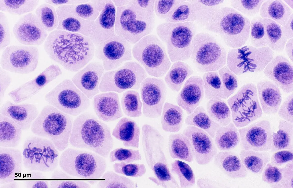
1.3.1 General introduction
Every cell comes from a cell that already existed. A cell grows, does its work, and at some point may divide. Cell division is the process in which one cell, the parent cell or mother cell, divides into two or more daughter cells. It is how life keeps going:
- In a unicellular organism, such as Amoeba or a bacterium, dividing is reproducing: one organism becomes two.
- In a multicellular organism, cell division turns the single-celled zygote into the whole body (growth), replaces worn-out and damaged cells (repair and regeneration), and makes the cells used in reproduction.
Before a cell can divide, it must copy everything a new cell needs, above all its DNA. Bacteria and other prokaryotes have a simple job: they copy their single circular chromosome, the two copies move apart as the cell grows, and the cell pinches in two. A eukaryotic cell has many long chromosomes inside a nucleus, so it divides in a series of distinct stages. Its division either keeps the chromosome number the same (a diploid cell gives two diploid daughter cells, genetically identical, for growth) or halves it (a diploid cell gives four haploid cells, for sexual reproduction).
Either way, a eukaryotic cell divides in two steps:
- Karyokinesis (Greek karyon, nucleus, and kinesis, movement): the division of the nucleus.
- Cytokinesis (kytos, a hollow vessel, a cell): the division of the cytoplasm, which makes two separate cells.
The division of the cytoplasm of a cell is called:
Pick an answer to see why.
1.3.2 Cell cycle
A cell that divides doesn’t spend most of its time dividing. It spends it growing and getting ready. The whole sequence of events from one division to the next, growth, copying of the DNA and division, is the cell cycle.
It helps to see the cell cycle as three cycles running together, each of which must finish before the cell can divide:
- The chromosome cycle. The DNA is copied, so that each chromosome becomes two identical sister chromatids; then the copies are shared out, one to each daughter cell.
- The cytoplasmic cycle. The cell grows, making more cytoplasm and organelles; then the cytoplasm divides.
- The centrosome cycle. In an animal cell the centrosome, which organizes the spindle, is copied; then the two centrosomes move apart to the two poles of the cell.
All this happens in two broad phases: interphase and the M phase.
1. Interphase
Interphase (Latin inter, between, and Greek phasis, appearance or stage) is the time between the end of one division and the start of the next. Under a light microscope the nucleus looks quiet: no chromosomes can be seen, only a faint network of chromatin. That is why interphase was once called the resting stage. But the cell is at its busiest, growing and making the molecules it needs for the next division. Interphase has three sub-phases:
G₁ phase (first gap, or first growth phase). The new cell grows. It makes RNA and proteins, including enzymes and structural proteins, carbohydrates and lipids, and new organelles, so it gets bigger. It also builds up the raw materials for copying DNA: nucleotides and energy-rich compounds such as ATP. The amount of DNA does not change in G₁. Near the end of G₁ the cell passes a check: is it large enough, is its DNA undamaged, are the outside signals right? Only then does it go on to copy its DNA.
S phase (synthesis phase). The cell copies (replicates) its DNA, and makes the histone proteins that the new DNA is wound round, and the proteins of the kinetochores. At the end of S each chromosome is made of two identical sister chromatids, joined at the centromere. The number of chromosomes is the same as before; the amount of DNA has doubled. In an animal cell the pair of centrioles is copied during S as well.
G₂ phase (second gap, or second growth phase). The cell keeps growing and makes the proteins needed for division, among them tubulin, the protein of the spindle fibers, and stores ATP. It finishes making new mitochondria and plastids, and the copied centrioles mature. It checks that all its DNA has been copied, and repairs any breaks made during copying, before it enters mitosis.
2. M phase
The M phase (mitotic phase) follows interphase. It is the division itself, in two parts:
- Karyokinesis, the division of the nucleus, which in mitosis has four stages: prophase, metaphase, anaphase and telophase. Each daughter nucleus gets the same, identical set of chromosomes.
- Cytokinesis, the division of the cytoplasm with its organelles, which makes two cells.
| Stage | Events within the cell |
|---|---|
| G₁ | The cell grows fast, making the structural and functional proteins it needs, and RNA, lipids and carbohydrates. |
| S | The DNA is replicated and histone proteins are made, so each chromosome comes to have two chromatids. |
| G₂ | RNA, proteins (tubulin for the spindle) and ATP are made; the copying of mitochondria, plastids and centrioles is completed. |
| M: karyokinesis | The nucleus divides, through prophase, metaphase, anaphase and telophase. |
| M: cytokinesis | The cytoplasm and organelles are shared between the two daughter cells: by a cleavage furrow in animal cells, by a cell plate in plant cells. |
G₀ phase
Not every cell keeps going round the cycle. A cell that stops dividing leaves the cycle from G₁ and enters a state called G₀ (G-zero, the quiescent stage). This happens when the signals that tell a cell to divide (mitogens, such as growth factors) are missing, or when the cell becomes specialized for a job. A cell in G₀ is not idle: it is fully active at its work, it simply doesn’t prepare to divide. Some cells stay in G₀ for life, such as most nerve cells and heart muscle cells. Others are reserve cells that can re-enter the cycle when needed: liver cells divide again if part of the liver is removed, and the cells round a wound start dividing to heal it. The step from G₁ into G₀ is linked to cell differentiation, the change of a cell into one kind with one job.
How long does a cycle take?
The length of the cell cycle depends on the kind of cell and on conditions such as temperature and food. Interphase always takes most of it. For a typical human cell growing in a dish (24 hours in all), G₁ takes about 46 % of the cycle, S about 33 %, G₂ about 17 % and M about 4 %. For many cells the shares are roughly: G₁ 30–40 %, S 30–50 %, G₂ 10–20 % and M 5–10 %.
In which phase of the cell cycle does each chromosome come to have two sister chromatids?
Pick an answer to see why.
A nerve cell that will never divide again is in:
Pick an answer to see why.
Measuring the phases: the mitotic index
You can’t time a cell in a fixed, stained slide: it is dead. But you can still find how long each stage lasts. In a growing root tip the cells divide independently, each at its own moment, so at any instant the cells are spread through the cycle. A stage that lasts twice as long catches twice as many cells. So the fraction of cells in a stage is the fraction of the cycle that stage takes:
The percentage of cells that are dividing (in any stage of mitosis) is the mitotic index:
1.3.3 Types of cell division
Cells divide in three ways:
- Direct cell division, or amitosis.
- Indirect cell division, or mitosis.
- Reductional cell division, or meiosis.
1. Direct cell division or amitosis
Amitosis (Greek a, without, and mitos, thread) is the simplest way for a nucleus to divide: it simply pinches in two, with no chromosomes coiling up into visible threads and no spindle (Figure 3.3).
- The nucleus gets longer, and a groove (constriction) appears round its middle.
- The groove deepens until the nucleus divides into two daughter nuclei.
- At the same time, or soon after, a groove forms in the cytoplasm too, and the cell divides into two roughly equal halves.
Because nothing makes sure that each daughter nucleus gets one copy of every chromosome, amitosis doesn’t share out the genes exactly. It is found where that matters little: the large macronucleus of Paramecium and other ciliates (which holds many copies of each gene) divides this way, and so do some cells of cartilage, of the mammalian liver, and of old or damaged tissues.
Bacteria, archaea and other simple organisms divide by binary fission: the cell copies its one circular chromosome, the two copies move apart as the cell grows longer, and the cell pinches in two, giving two identical cells. Like amitosis, it has no spindle and no condensed chromosomes, so many books treat the two together. Amoeba and Paramecium also reproduce by binary fission, splitting into two; but their nucleus (the micronucleus, in Paramecium) divides by mitosis, with a spindle.
2. Indirect cell division or mitosis
Mitosis (Greek mitos, thread) is the division in which one cell gives two daughter cells with the same number and kind of chromosomes as the mother cell. A human body cell with 46 chromosomes gives two cells with the same 46; an onion root cell with 16 gives two with 16. Because the number stays equal, it is also called equational division, and because the nucleus goes through visible stages before dividing, indirect division.
Mitosis happens wherever an organism grows, repairs itself or reproduces asexually: in the somatic (body) cells of animals, in the meristems of plants (the tips of roots and shoots, and the cambium), in the early embryo, and in the germ cells of the testes and ovaries, which multiply by mitosis before some of them go on to meiosis.
Mitotic cell division has two parts, after interphase:
- A. Interphase
- B. Mitotic phase: 1. karyokinesis, 2. cytokinesis
A. Interphase
Interphase is the stage between two divisions. The cell looks inactive but is metabolically very active:
- The chromosomes are long, thin, uncoiled chromatin fibers, spread through the nucleus; a nucleolus and the nuclear envelope are clearly seen.
- Each chromosome makes an exact copy of itself (in S phase).
- Proteins and RNA are made.
- Organelles such as mitochondria, plastids and centrioles are copied.
B. Mitotic phase: 1. Karyokinesis
Mitosis is one smooth process, but it is described in four stages. Step through them in the widget, then read what happens in each.
a. Prophase (Greek pro, first or before, and phasis, stage) is the first visible stage of nuclear division, and usually the longest stage of mitosis.
- The nucleus becomes rounder, and the cytoplasm becomes thicker (more viscous).
- The long chromatin fibers coil and condense into chromosomes, which become shorter and thicker. Proteins called condensins fold them up.
- Each chromosome can now be seen to be double: two sister chromatids joined at the centromere.
- In an animal cell the two centrosomes (each with a pair of centrioles) move towards opposite ends of the cell, the poles. Each sends out short microtubules that radiate like the rays of a star, the astral rays; a centrosome with its rays is an aster. Between the two asters the spindle starts to form.
- By the end of prophase the nucleolus and the nuclear envelope break up and disappear, and the asters lie where the poles of the spindle will be.
b. Metaphase (Greek meta, after) comes after the chromosomes are free in the cell.
- Spindle fibers from each pole attach to the chromosomes at their centromeres, on two small discs of protein called kinetochores, one on each sister chromatid. One chromatid is held by fibers from one pole, its sister by fibers from the other.
- Pulled from both sides, the chromosomes move to the middle of the cell. This movement to the equator is called congression.
- They line up in a single plane across the cell, the equatorial plate or metaphase plate: their centromeres on the plate, their arms pointing any way. In a flat view across the plate, the smaller chromosomes tend to lie towards the middle and the larger ones round the edge.
- The chromosomes are now at their shortest and thickest, which is why chromosomes are counted and photographed at metaphase.
c. Anaphase (Greek ana, up or back) is the shortest stage, and the stage of migration.
- The centromere of each chromosome splits, so the two sister chromatids come apart. Each is now a daughter chromosome of a single chromatid.
- The daughter chromosomes move to opposite poles, centromere first. The spindle fibers attached to them shorten (their microtubules are taken apart at the ends), while the fibers that run between the two groups lengthen and push the poles apart.
- Dragged by its centromere, each chromosome takes the shape of a letter that depends on where its centromere is: a V for a metacentric chromosome, an L for a submetacentric, a J for an acrocentric and an I (a straight rod) for a telocentric (Figure 3.5).
- By the end of anaphase a complete, identical set of chromosomes has reached each pole.
d. Telophase (Greek telos, end) is the stage of reorganization, prophase in reverse.
- The daughter chromosomes reach the poles. They take up water and uncoil (despiralize), becoming long, thin chromatin fibers again.
- The spindle fibers disappear.
- A nuclear envelope forms round each group of chromosomes, and a nucleolus appears in each.
- By the end of telophase the cell has two daughter nuclei, one at each pole, with identical chromosomes.
At which stage of mitosis do the chromosomes line up on the equator of the spindle?
Pick an answer to see why.
During anaphase, an acrocentric chromosome looks like the letter:
Pick an answer to see why.
B. Mitotic phase: 2. Cytokinesis
Cytokinesis divides the cytoplasm, with its organelles, between the two new nuclei. It usually begins in late anaphase or telophase. Animal and plant cells do it in opposite ways (Figure 3.6).
a. Cell plate formation (in plant cells). A plant cell can’t pinch in: its rigid wall is in the way. So it builds a new wall across its middle, from the inside out. Between the two daughter nuclei, the leftover spindle microtubules form a barrel-shaped framework called the phragmoplast. Small vesicles from the Golgi bodies (with some from the ER), filled with pectin and other wall materials, travel along it to the equator, where they gather and fuse into a flat disc, the cell plate. The plate grows from the center outward until it meets the old wall. It becomes the middle lamella, and each daughter cell lays down its own primary wall on its side.
b. Furrowing (in animal cells). A ring of protein filaments, actin and myosin (the same proteins that make muscles contract), forms just under the cell membrane round the cell’s equator. As the ring tightens, it pulls the membrane in, making a groove, the cleavage furrow, which deepens from the outside in. When its edges meet in the middle, the cell is pinched into two daughter cells. Bacteria divide with a ring too, though of a different protein (FtsZ, related to tubulin), which pinches the cell in while a wall (a septum) is laid down across it, and many fungi lay down a cross-wall in the same way.
The result is two daughter cells, each with a nucleus holding the same chromosomes as the mother cell and a share of its cytoplasm. Each enters G₁, and the cycle starts again.
In which direction does the cell plate of a dividing plant cell grow?
Pick an answer to see why.
Significance of mitosis
- Growth. Mitosis builds a multicellular organism from a single zygote into an adult, every cell with the same chromosomes.
- Asexual reproduction. Many organisms reproduce by mitosis alone: yeast by budding, Hydra by buds, plants by fragments, runners, tubers and stem cuttings.
- Genetic stability. Each daughter cell gets the same number of chromosomes as the mother cell, so the number stays constant from cell to cell.
- Repair, regeneration and healing. Mitosis replaces worn-out and damaged cells: new skin over a cut, new blood cells (about two million red blood cells every second), a lizard’s regrown tail.
- Keeping cells at a workable size. A cell that grows too big has too little surface for its volume, and too much cytoplasm for its one nucleus to run; division restores a workable size.
- Genetic consistency. The daughter cells are the same as the mother cell in both the number (quantity) and the kind (quality) of their genes.
- When it goes wrong: cancer. When the controls of the cell cycle fail, cells divide without stopping and form a lump, a tumor. Uncontrolled mitosis is what makes cancer grow.
3. Reductional cell division or meiosis
Meiosis (Greek meioun, to make smaller, and -osis, a process) is the division in which a diploid mother cell (2n) divides into four haploid daughter cells (n), each with half the number of chromosomes. Because it halves the number, it is also called reductional division. Halving matters because at fertilization two gametes join: if each carried a full 2n, the number would double every generation.
Meiosis happens only in special cells, the meiocytes, in organs of sexual reproduction: the spermatocytes in the testes and oocytes in the ovaries of animals, and the pollen mother cells (microspore mother cells) in anthers and the megaspore mother cells in ovules of flowering plants.
Like mitosis, meiosis comes after an interphase in which the DNA is copied, so each chromosome enters meiosis with two sister chromatids. Its G₂ is short or absent. Then come two divisions one after the other, meiosis I and meiosis II, with no copying of DNA between them. The short gap between the two, if there is one, is called interkinesis.
Meiosis I
Meiosis I halves the chromosome number: it gives two cells, each with half as many chromosomes as the mother cell. It is divided into karyokinesis I and cytokinesis I.
A. Karyokinesis I
Karyokinesis I has four phases: prophase I, metaphase I, anaphase I and telophase I.
Prophase I is long and complicated, the longest phase of meiosis. In it, each chromosome finds its partner, the homologous chromosome from the other parent, and the two exchange pieces. It is described in five sub-phases (Figure 3.7):
i. Leptotene or leptonema (Greek leptos, thin, and tainia or nema, band or thread), the thin-thread stage:
- The nucleus enlarges.
- The chromosomes begin to condense and become visible as long, thin, single-looking threads. Along them lie darker, bead-like thickenings, the chromomeres. (Each chromosome has already been copied, but its two chromatids lie so close that they can’t yet be told apart.)
- In many cells the ends of the chromosomes gather on one side of the nucleus, near the centrosome, in a loop-like cluster called the bouquet stage.
- The nuclear envelope and the nucleolus remain.
ii. Zygotene or zygonema (Greek zygon, yoke), the paired-thread stage:
- The chromosomes get shorter and thicker, and each finds its homologous partner, the chromosome of the same length and shape from the other parent.
- The two homologs pair up side by side, gene against matching gene, like a zip closing. This pairing is called synapsis, and it is held together by a ladder of proteins between them, the synaptonemal complex.
- Each paired couple is called a bivalent (two chromosomes). A human cell forms 23 bivalents.
- In an animal cell the two asters keep moving apart.
iii. Pachytene or pachynema (Greek pachys, thick), the thick-thread stage, which lasts much longer than leptotene and zygotene:
- The bivalents become shorter and thicker.
- The two chromatids of each chromosome become visible, except at the centromere where they are still joined. So each bivalent is seen to have four chromatids, and is also called a tetrad. The two chromatids of the same chromosome are sister chromatids; a chromatid of one homolog and a chromatid of the other are non-sister chromatids.
- Crossing over happens: two non-sister chromatids break at matching points and swap the pieces beyond the break, so each comes to carry part of the other parent’s chromosome.
iv. Diplotene or diplonema (Greek diplous, double):
- The synaptonemal complex comes apart, and the two homologs of each bivalent begin to separate, except at the points where crossing over happened. There the chromatids stay crossed, in X-shaped joins called chiasmata (singular chiasma, Greek for a cross like the letter χ).
- The nuclear envelope is still there; the nucleolus gets smaller.
- In the oocytes of many animals, including humans, meiosis pauses at diplotene, sometimes for years.
v. Diakinesis (Greek dia, across, and kinesis, movement), the last sub-phase:
- The chromosomes are at their most condensed, and the four chromatids of each tetrad are clearly visible. The two chromatids of each chromosome are still joined at the centromere.
- The bivalents move apart from one another, towards the edge of the nucleus.
- The chiasmata slide towards the ends of the chromosomes: terminalization.
- The nucleolus and the nuclear envelope disappear, and the spindle starts to form.
Metaphase I. The nuclear envelope and the nucleolus have gone, and the spindle is complete.
- The bivalents (not single chromosomes) move to the equator and line up there. The two homologs of each bivalent lie on either side of the equator, so that the bivalents form two parallel rows, two equatorial plates.
- Each homolog’s centromere is attached to spindle fibers from one pole only: the two centromeres of a bivalent face opposite poles, equally far from the equator. The two sister chromatids of a chromosome act as one, facing the same pole.
- The chromosomes are still joined in bivalents by their chiasmata.
- Which homolog of a pair faces which pole is a matter of chance, and each pair lines up independently of every other (Figure 3.8).
Anaphase I comes after metaphase I.
- The centromeres do not split: the two chromatids of each chromosome stay joined.
- The two homologous chromosomes of each bivalent separate and move to opposite poles, as the chiasmata come undone and the spindle fibers shorten. This separation of homologs is called disjunction.
- Of each pair, one chromosome goes to each pole: which one, the mother’s (maternal) or the father’s (paternal), is decided by how the pair lined up at metaphase I. A pole receives a mixture of maternal and paternal chromosomes.
- By the end of anaphase I there are two groups of chromosomes, each with half the number in the mother cell, though each chromosome still has two chromatids.
Telophase I
- Each pole has one set of chromosomes, each of two chromatids. The chromosomes may uncoil a little into chromatin threads.
- The spindle fibers disappear.
- A nuclear envelope and a nucleolus may form round each set, giving two haploid nuclei. (In some organisms the cell goes straight from anaphase I into meiosis II, with no telophase I.)
B. Cytokinesis I
Not every cell divides its cytoplasm at this point. When it does, animal cells form a cleavage furrow and plant cells a cell plate, giving two daughter cells, each with half the chromosome number of the mother cell. In the pollen mother cells of many flowering plants (most dicots), the cytoplasm divides only once, at the very end of meiosis II, giving all four cells at once.
Interkinesis
Interkinesis is the short pause, if any, between meiosis I and meiosis II. The cell may make some proteins and RNA, but it has no S phase: the DNA is not copied again. This is the key to meiosis: one copying of the DNA, two divisions, so the DNA per cell ends at half. In some organisms interkinesis is very short or missing.
Meiosis II
Meiosis II is much like mitosis, in each of the two haploid cells from meiosis I. It separates the two sister chromatids of each chromosome. The number of chromosomes per cell is the same before and after it (n to n), so it is also called equational division. It has karyokinesis II and cytokinesis II.
A. Karyokinesis II
- Prophase II. The chromosomes, each still of two chromatids joined at the centromere, condense again and become shorter, thicker and clearly visible. The nuclear envelope and the nucleolus (if they formed) disappear, and a new spindle forms, usually at right angles to the spindle of meiosis I.
- Metaphase II. The chromosomes line up singly on the equator of the new spindle. Each chromosome’s centromere is attached to spindle fibers from both poles: one chromatid faces one pole, its sister the other, just as in mitosis.
- Anaphase II. The centromere of each chromosome splits, and the sister chromatids separate and move to opposite poles, pulled by the shortening spindle fibers while the fibers between the groups lengthen. Once apart, each chromatid is called a chromosome.
- Telophase II. The chromosomes reach the poles, uncoil and stretch out into chromatin fibers again; the spindle disappears; a nuclear envelope forms round each group, and a nucleolus reappears. There are now four haploid nuclei.
B. Cytokinesis II
The cytoplasm divides again, by a cell plate in plant cells or a cleavage furrow in animal cells. The result: from one diploid mother cell, four haploid cells, each with half the chromosome number and half the DNA of the mother cell in G₁, and no two alike.
Crossing over takes place between:
Pick an answer to see why.
In which phase do the homologous chromosomes separate and move to opposite poles?
Pick an answer to see why.
How many times is the DNA copied during one complete meiosis?
Pick an answer to see why.
Importance of meiosis
- It makes gametes and spores. Meiosis forms the haploid gametes of animals (sperm and eggs) and the haploid spores of plants, which sexual reproduction needs.
- It keeps the chromosome number constant from one generation to the next. Meiosis halves the number in the gametes (n), and fertilization restores it (n + n = 2n), so the species keeps its genetic balance.
- It causes genetic variation, in two ways: crossing over, the exchange of pieces between non-sister chromatids of homologous chromosomes, makes new combinations of genes on each chromosome; and independent assortment, the random way each pair lines up at metaphase I, makes new combinations of whole chromosomes in each gamete.
- It segregates genes. The two copies of each gene, on the two homologs, are separated into different gametes (Mendel’s law of segregation), and pairs on different chromosomes are sorted independently (his law of independent assortment). Meiosis is the reason Mendel’s laws work.
- It can give rise to polyploids. If the chromosomes fail to separate (non-disjunction) in every pair, a gamete may get two full sets (2n) instead of one; joining a normal gamete, it gives a triploid, or with another 2n gamete a tetraploid, plant. Many crops arose this way: bread wheat has six sets (2n = 42), the potato four (2n = 48).
- It gives the variation that breeders select. The many small differences between offspring (continuous variation) are what plant and animal breeders choose from to improve crops and livestock.
- It shows how organisms are related. Meiosis runs in essentially the same way in all sexually reproducing organisms, from yeast to rhododendrons to people, a sign that they share a common ancestor.
- It makes alternation of generations possible. In plants the life cycle alternates between a haploid generation (n) and a diploid generation (2n); meiosis is the step from 2n to n.
- It keeps the reproductive cycle regular, each generation passing through the same steps: 2n by fertilization, n by meiosis.
Mitosis and meiosis compared
| Feature | Mitosis | Meiosis |
|---|---|---|
| Where | Body (somatic) cells, meristems, and germ cells while they multiply. | Meiocytes in the gonads (testes, ovaries) and in the anthers and ovules of plants. |
| Divisions | One. | Two (meiosis I and II), after one copying of the DNA. |
| Prophase | Short and simple, with no sub-phases. | Prophase I is long, with five sub-phases: leptotene, zygotene, pachytene, diplotene and diakinesis. |
| Pairing | Homologous chromosomes do not pair; no crossing over, no chiasmata. | Homologs pair (synapsis) into bivalents; crossing over; chiasmata. |
| Metaphase | Single chromosomes, each of two chromatids, line up on one plate. | At metaphase I, bivalents of four chromatids (tetrads) line up, in two rows. |
| Anaphase | Centromeres split; sister chromatids go to opposite poles. | At anaphase I, whole homologs go to opposite poles (centromeres don’t split); at anaphase II, sister chromatids separate. |
| Telophase | Always happens. | Telophase I is short or may be missed. |
| Interkinesis | None. | A short pause between meiosis I and II, with no copying of DNA. |
| Daughter cells | Two, diploid (2n) like the mother cell, with the same DNA as the mother cell in G₁. | Four, haploid (n), each with half the DNA of the mother cell in G₁. |
| Genetic make-up | Identical to the mother cell and to each other. | Different from the mother cell and from each other. |
| Role | Growth, repair, regeneration, asexual reproduction. | Gametes and spores for sexual reproduction; variation. |
Exercise
A. Tick the correct alternatives
- Which of the following is a key difference between mitosis and meiosis?
Pick an answer to see why.
- What is the main purpose of the cell cycle?
Pick an answer to see why.
- Which of these lists the phases of mitotic cell division, with the interphase before it?
Pick an answer to see why.
- What is a characteristic feature of mitotic cell division?
Pick an answer to see why.
- What is the role of centrioles in cell division?
Pick an answer to see why.
- How would a change in the rate of cell division most directly affect an organism’s growth and development?
Pick an answer to see why.
B. Answer the following questions
Define cell cycle.
Answer
What is mitotic cell division?
Answer
What is meiosis?
Answer
Name the phases of cell division.
Answer
What is the difference between mitotic and meiotic cell division?
Answer
What is the significance of mitotic cell division?
Answer
Explain the difference between mitosis and meiosis in terms of the number of daughter cells and their genetic composition.
Answer
How can one identify the phase of cell division through microscopic study?
Answer
Describe the phases and sub-phases of cell division in detail.
Answer
Compare the difference between mitotic and meiotic cell division in terms of their roles in an organism.
Answer
Explain the significance of meiotic cell division.
Answer
How can understanding the difference between mitosis and meiosis be applied in the field of genetics?
Answer
How can identifying the phase of cell division be useful in medical diagnosis and treatment?
Answer
How can knowledge of the phases and sub-phases of cell division be applied in the field of cell biology?
Answer
How can understanding the difference between mitotic and meiotic cell division be applied in the field of reproductive biology?
Answer
How can understanding the significance of mitotic and meiotic cell division be applied in the field of medicine?
Answer
Analyze the role of mitosis and meiosis in the life cycle of a sexually reproducing organism.
Answer
Evaluate the importance of being able to identify the phase of cell division in the context of cancer research.
Answer
Compare and contrast the roles of mitotic and meiotic cell division in the context of human health and disease.
Answer
Printable PDFs
- Study notes 4 pages
- Worksheet 5 pages Answer key
Quiz yourself: Living Things and Their Structure, Grades 5–12 →
Practice every question in the book, and keep track of the ones you miss →
A companion to your textbook, not a replacement for it: study from the book your school uses, and where the two differ, follow your teacher. Found a mistake? Please tell us.