
Right now, while you read this, waves are passing through your body. Radio waves from FM stations in Kathmandu, Pokhara and Biratnagar. Microwaves carrying messages to and from every phone around you, and from the Wi-Fi router. Infrared from the warm walls of the room. Light from the window, and sound from the street. You can’t feel most of them, yet you use them all day.
In this unit you’ll find out what a wave is (it is not what most people think), the two ways a wave can shake, and the whole family of electromagnetic waves, from radio to gamma rays. Then you’ll see how echoes measure the depth of the sea, how bats hunt in the dark, and how doctors look inside your body with sound, X-rays and a spinning X-ray machine called a CT scanner.
Waves all around us
We use the energy carried by many kinds of waves:
- light and heat (infrared) from the Sun;
- radio waves from FM radio and TV transmitters;
- microwaves for mobile phones, Wi-Fi and microwave ovens;
- infrared from a TV remote control;
- X-rays and ultrasound (sound too high-pitched to hear) to look inside the human body;
- and networks of waves for security checks, measuring distances, and much more.
These waves differ in one important way: how fast they shake. Radio waves shake slowly (they have a low frequency); light, X-rays and gamma rays shake extremely fast (a high frequency). You’ll meet them all in order below.
Wave
Activity 10.1: what moves in a wave?
Here is what happens. When your finger taps the water, it pushes the water particles near it down and up. Each particle is joined to its neighbors, so as it moves, it pulls and pushes them, and they start moving up and down too, a moment later. They pass the motion on to their neighbors, and so on. So the disturbance spreads outwards across the surface as a ripple, carrying energy with it. But each particle only oscillates (moves to and fro) about its own resting place, called its mean position. The paper boat shows this: it rides up and down on the particles under it, but stays where it is.
The rope works the same way. Your hand moves the end of the rope up and down. That end pulls the next part of the rope with it, which pulls the next part, a little later each time. Some parts of the rope are raised while others are pushed down, and the shape travels along the rope. The ribbon shows that each bit of rope only moves up and down about its place, while the humps move forward.
So a wave is a periodic disturbance that carries energy from one place to another, without carrying the material along with it. (“Periodic” means it repeats again and again.)
Not every wave needs a material to travel in. In light, radio waves and X-rays, what goes up and down is electric and magnetic fields, and these waves carry energy across empty space.
A leaf floats on a pond. A ripple passes under it. What does the leaf do?
Pick an answer to see why.
Types of waves
Waves are sorted in two ways:
- by whether they need a medium (a material to travel through): mechanical waves need one; electromagnetic waves don’t;
- by which way the particles move: in longitudinal waves they move back and forth along the direction the wave travels; in transverse waves they move at right angles to it.
| Types of waves | ||
|---|---|---|
| Mechanical waves (need a medium) | longitudinal, e.g. sound | transverse, e.g. a wave on a rope |
| Electromagnetic waves (need no medium) | always transverse, e.g. light, radio waves |
Transverse waves and longitudinal waves
Activity 10.2: two ways to shake a slinky
Longitudinal wave
When you push and pull the end of the slinky steadily, the rings near your hand are pushed close together: a compression. Then, as you pull back, they spread apart: a rarefaction. This repeats again and again, and the compressions and rarefactions travel along the slinky.
In a longitudinal wave, the particles of the medium vibrate back and forth in the same direction as the wave travels. Sound is a longitudinal wave. Longitudinal waves can travel through all three states of matter: solids, liquids and gases.
Transverse wave
When you move the end of the slinky from side to side, the rings move left and right, while the humps travel forward along the slinky.
In a transverse wave, the particles of the medium vibrate at right angles (perpendicular) to the direction in which the wave travels. A wave on a rope or a string, and the ripples on the surface of water, are transverse waves. Mechanical transverse waves can travel through solids and along the surface of liquids, but not through gases: the particles of a gas aren’t held to one another, so when one moves sideways, it doesn’t drag its neighbors with it.
Words used for waves
- Compression: in a longitudinal wave, the part where the particles are crowded together (the pressure is higher).
- Rarefaction: in a longitudinal wave, the part where the particles are spread apart (the pressure is lower).
- Crest: in a transverse wave, the highest part, raised above the mean position.
- Trough: in a transverse wave, the lowest part, below the mean position.
- Amplitude (A): the largest distance a particle moves from its mean position: from the mean position to the top of a crest, or to the bottom of a trough. A big amplitude means a loud sound, or a high water wave.
- Wavelength (λ, the Greek letter lambda): the length of one complete wave. In a transverse wave, the distance from one crest to the next crest (or one trough to the next trough). In a longitudinal wave, the distance from the middle of one compression to the middle of the next (or one rarefaction to the next). Its SI unit is the meter (m).
- Frequency (f): the number of complete waves produced (or passing a point) in one second. Its SI unit is the hertz (Hz): 1 Hz is one wave per second.
- Time period (T): the time taken to produce one complete wave.
If one complete wave is produced in T seconds, then f waves are produced in one second, so
Wave velocity
Not all waves travel equally fast. How fast a wave travels depends on the medium, the kind of wave, and so on. The distance a wave travels in one second is its wave velocity (v), in m/s.
Why is it equal to frequency × wavelength? In one second, the source makes f waves. Each wave is λ long. So in one second, the front of the wave moves forward by f lengths of λ:
A wave’s frequency is doubled, but it travels in the same medium, so its speed stays the same. What happens to its wavelength?
Pick an answer to see why.
Mechanical waves and electromagnetic waves
Shout in an empty valley and the echo comes back. Shout on the Moon, where there is no air, and nobody would hear you, not even someone standing next to you. Yet sunlight crosses 150 million km of empty space to reach us. Some waves need a medium to travel through; others don’t.
Mechanical waves
A wave that needs a medium to travel (propagate) through is a mechanical wave. Sound is a mechanical wave. A mechanical wave is energy passed along by the vibration of the particles of a material; that material (solid, liquid or gas) is its medium.
- As it travels, the medium absorbs some of its energy, so the wave grows weaker. A mechanical wave can only travel a limited distance.
- Its speed is different in different media. Under normal conditions, sound travels at about 343 m/s in air, 1,481 m/s in water and 5,120 m/s in iron.
- Mechanical waves may be longitudinal (sound) or transverse (a wave on a rope).
Electromagnetic waves
The light of the Sun and the stars reaches the Earth through the vacuum of space. A wave that doesn’t need a medium to travel is an electromagnetic wave. Light, ultraviolet rays, the infrared rays that carry heat, radio waves and X-rays are electromagnetic waves. Passing energy in the form of electromagnetic waves is called radiation.
- In a vacuum, nothing absorbs their energy, so they can travel enormous distances: light from galaxies billions of light-years away still reaches us.
- In a vacuum they all travel at the speed of light, 3 × 10⁸ m/s. Light from the Sun reaches the Earth in about 8 minutes 20 seconds.
- They are transverse waves: the electric and magnetic fields vibrate at right angles to the direction the wave travels.
- They are made naturally (by the Sun and stars) and artificially (by radio transmitters, lamps, X-ray machines).
Why can astronauts on the Moon talk to each other only by radio, even when standing side by side?
Pick an answer to see why.
Electromagnetic spectrum
Activity 10.3
The light from an electric lamp and the signal from a radio station are both electromagnetic waves. They travel at the same speed, but have very different frequencies and wavelengths. The whole range of electromagnetic waves, from the lowest frequency to the highest, is called the electromagnetic spectrum. In order of decreasing wavelength (increasing frequency), its parts are:
radio waves → microwaves → infrared rays → visible light → ultraviolet rays → X-rays → gamma rays
- At one end, radio waves have the longest wavelength and the lowest frequency.
- At the other end, gamma rays have the shortest wavelength and the highest frequency.
- The shorter the wavelength (the higher the frequency), the more energy the wave carries. That is why X-rays and gamma rays can damage living cells, while radio waves can’t.
Applications of electromagnetic waves
Radio waves
Wavelength: from about 30 cm to many kilometers. Radio waves don’t harm living cells, so they are safe for living beings.
- A transmitter produces radio waves that carry the signals of radio and TV stations.
- A radio telescope, a big dish, catches the radio waves given out by objects in space, to study the universe.
Microwaves
Wavelength: from about 1 mm to 30 cm.
- In a microwave oven, microwaves make the water molecules in food vibrate and spin very fast; the food heats up from the inside, and is cooked.
- Radar uses microwaves: an airport surveillance radar sends out microwaves and catches their reflections from airplanes, to control air traffic. The police’s radar speed gun measures the speed of vehicles the same way.
- Microwaves carry signals in satellite communication, mobile phones, Wi-Fi and Bluetooth.
Infrared rays
“Infra” means below: infrared is “below red”. Its frequency is lower than that of red light, and its wavelength longer: from about 7.5 × 10⁻⁷ m to 10⁻³ m. When strong infrared falls on our body, we feel it as heat. Every warm object gives it out; you give it out too.
- Heaters and some cookers heat things with infrared rays.
- A radiation thermometer (the kind pointed at your forehead) measures body temperature from the infrared your body gives out.
- Infrared CCTV cameras and thermal imaging cameras see in the dark, to watch for activity at night.
- Remote controls send infrared signals to switch a TV on and off, and to steer toy cars and drones.
- Astronomers study the infrared given out by objects in space, which passes through clouds of dust that block light.
Visible light
The retina of the eye can detect only this narrow band of electromagnetic waves. Red light has the longest wavelength (about 7.5 × 10⁻⁷ m, or 750 nm), and violet light the shortest (about 4 × 10⁻⁷ m, or 400 nm). Light from a source strikes an object and is reflected; when the reflected light enters our eyes, we see the object. A leaf looks green because it absorbs most of the other colors of sunlight and reflects green.
- We use it to see.
- Plants absorb visible light for photosynthesis.
- Screens of mobile phones, televisions and computers give out visible light.
- Signals for communication travel as light through optical fibers (mostly infrared, just beyond red, which glass absorbs the least).
Ultraviolet rays
“Ultra” means beyond: ultraviolet (UV) is “beyond violet”. Wavelength: from about 4 × 10⁻⁷ m down to 10⁻⁸ m. We can’t see UV, but long exposure to it burns the skin, and too much can cause skin cancer. The Sun is the main source of UV on the Earth; UV tubes also make it.
- In a CFL or tube light, the gas inside gives out UV; the fluorescent powder coating the inside of the glass absorbs it and glows with visible light.
- UV kills microbes: food, clothes and utensils can be disinfected by putting them in direct sunlight.
- In SODIS (solar disinfection), clear plastic bottles of water are left in direct sunlight for at least six hours; the UV (with the heat) kills the germs, making the water safe to drink.
- Our skin makes vitamin D when UV from the Sun falls on it. That is why babies in Nepal are given a massage in the morning sun.
X-rays
Wavelength: about 10⁻⁸ m to 10⁻¹¹ m. X-rays reach the Earth’s upper atmosphere from space (the air stops them), and are made artificially in X-ray machines.
- X-ray photography and CT scans show broken bones and injuries inside the body.
- X-ray security scanners at airports look for hidden dangerous objects in luggage.
- In X-ray therapy, powerful beams of X-rays are aimed at cancer cells to destroy them.
- In research, X-rays reveal how atoms are arranged in crystals and molecules (this is how the shape of DNA was found), and help identify minerals.
Gamma rays
Gamma rays (γ-rays) are given out by radioactive substances, whose unstable nuclei break up and release energy. They have the highest frequency and shortest wavelength of all, and carry the most energy. They are dangerous: they destroy living cells and can cause cancer. But used with care:
- a controlled beam of gamma rays destroys cancer cells: this treatment is called radiotherapy;
- they sterilize surgical instruments (kill every microbe on them);
- they sterilize food, so that it keeps for a long time;
- in nuclear medicine, a substance that gives out gamma rays (a tracer) is put into the body, and a camera follows the rays to see how an organ is working.
Which electromagnetic wave does a TV remote control use?
Pick an answer to see why.
Which of these lists is in order of increasing frequency?
Pick an answer to see why.
Reflection of sound
Sound travels out from its source through any medium. When it strikes a hard surface, it bounces back. Sound that comes back after striking a hard surface is called reflected sound. The echo you hear when you shout in front of a tall cliff or a big wall is reflected sound.
You hear an echo separately only if it comes back at least about 0.1 s after the original sound; sooner, and the two blur together. In 0.1 s, sound in air goes about 34 m, there and back, so the wall must be at least about 17 m away.
Uses of reflected sound
In nature. Bats give out very high-pitched sounds (ultrasound) and listen to the echoes from things nearby. From the echoes they find obstacles and their prey, even in total darkness. Finding the way by reflected sound is called echolocation. Dolphins use echolocation in murky water too.

The ultrasonic ruler. An ultrasonic ruler (distance meter) has a sender that sends out a pulse of ultrasound towards an object, and a receiver that catches the echo. It measures the time between sending and receiving, and works out the distance from it, as you did with the cliff. Builders use one to measure rooms.
SONAR. SONAR (sound navigation and ranging) is the technology of finding distances by sending out ultrasound from an instrument and timing how long its echo takes to return from an object. A fathometer (echo sounder) on a ship uses SONAR to measure the depth of the sea: it sends a pulse of ultrasound straight down and times the echo from the sea bed. Fishermen use it to find shoals of fish. Ultrasonography in hospitals works on the same idea, with echoes from inside the body.
Ship: traced from the Historic American Engineering Record (HAER AK-49), public domain
Security and safety. Alarms can be built on reflected ultrasound: burglar alarms on the doors of houses, which sound when someone moves in front of them and changes the echo; and the parking sensors in a car’s bumper, which beep faster as the echo from an obstacle comes back sooner.
Acoustic protection
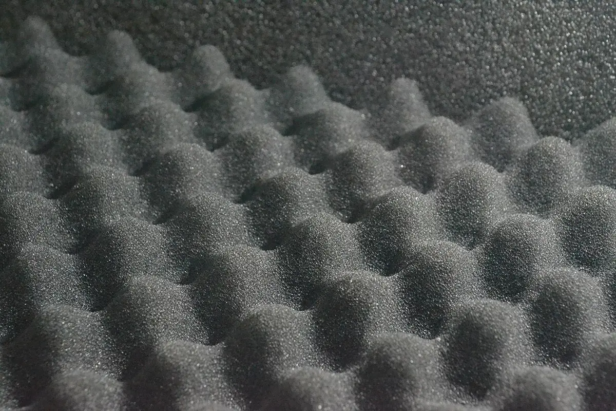
In an empty room with bare walls, your voice sounds booming and unclear, because the sound bounces back and forth off the walls many times. Smooth, hard walls reflect sound well; soft, porous materials (foam, curtains, carpets, cloth) absorb it.
Acoustic protection is the use of soft, porous materials on walls (and ceilings and floors) to absorb unwanted sound, so that it isn’t reflected. It is used in cinema halls, music rooms, recording studios, seminar halls and auditoriums, so that speech and music sound clear. Walls can be made rough, or covered with foam boards or panels full of small holes. The studios of Radio Nepal are built this way.
Rohan stands in front of his neighbor’s house and claps. He hears the echo 0.4 s later. Sound travels at 340 m/s. How far away is the house?
Pick an answer to see why.
Use of waves in diagnosing the body
Activity 10.4
Ultrasonography
Ultrasonography (USG) uses ultrasound to look at the organs inside the body.
- A hand-held part called the transducer (the probe) is pressed on the skin, with a little gel to keep out air. It sends ultrasound of very high frequency (usually 2 to 15 million Hz) into the body.
- Each time the ultrasound meets a boundary between two kinds of tissue (between fluid and muscle, or muscle and bone), part of it is reflected back.
- The same transducer receives the echoes.
- A computer measures how strong each echo is and how long it took to come back (the longer the time, the deeper the boundary), and builds a picture of the inside of the body from them. The picture is called a sonogram.
Ultrasound doesn’t harm the cells of the body. It shows soft tissues and body fluids well. It is used to check:
- stones in the kidneys and gall bladder;
- tumors in the brain and pancreas (in a baby’s brain, through the soft spot of the skull);
- diseases of the liver, the intestines and the uterus;
- the size, health and movements of a baby in the womb (fetus), and any abnormality. 3D ultrasound shows the baby even more clearly.
Ultrasound used to check the heart (its beating, and how its valves open and close) is called echocardiography.
X-ray imaging
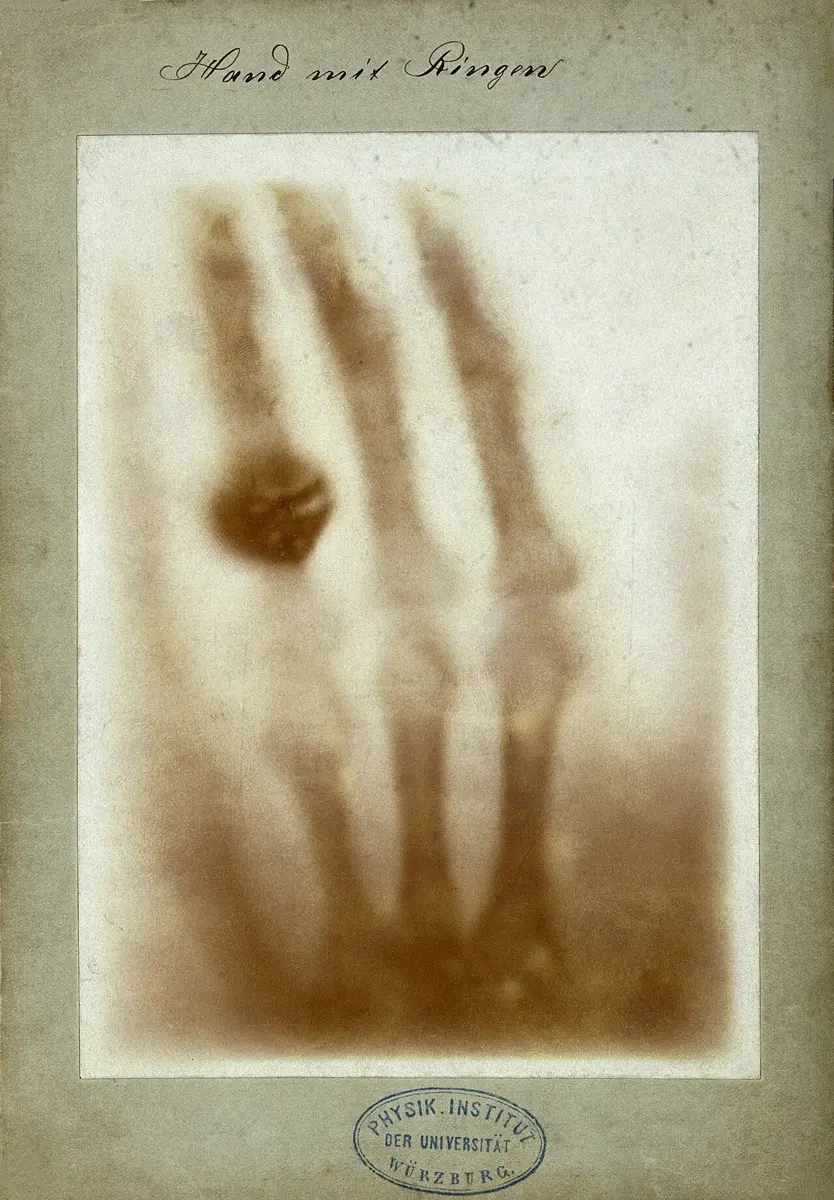
X-ray imaging uses X-rays to make pictures of the inside of the body for diagnosis; an X-ray photograph is simply called an X-ray. The machine sends a beam of X-rays through the body, and the picture is made from the shadows the body casts.
- The patient stands or lies in front of the X-ray machine (the X-ray tube).
- When the machine is switched on, X-rays pass through the body and fall on an X-ray film (or, in modern machines, an electronic detector) behind it.
- Different tissues absorb different amounts of X-rays. Bones absorb the most, so few X-rays reach the film behind them: their shadows come out white. Muscle and fat absorb less: they make faint, gray shadows. Air-filled organs like the lungs let almost all the X-rays through: they look black.
- A modern detector sends the picture to a computer screen, and it can be printed.
X-rays are used to check broken bones, the roots of teeth, infections (such as pneumonia and tuberculosis in the lungs), tumors and blocked arteries.
X-ray: Mikael Häggström, CC0, via Wikimedia Commons
Safety. Strong X-rays are harmful: too much exposure damages cells, can cause genetic changes, and increases the risk of cancer. When an X-ray machine is on, some X-rays scatter around the room, so the staff stand behind a screen, and lead aprons are used to cover the parts of the body not being X-rayed. (A single X-ray photo of the chest gives only a very small dose, about as much as ten days of the natural radiation all around us.)
CT scan
A CT scan (computerized tomography scan) is an advanced kind of X-ray imaging. An ordinary X-ray squashes everything into one flat shadow: a bone in front hides whatever is behind it. A CT scan instead makes pictures of thin slices of the body.
- The patient lies on a table that moves slowly into a big doughnut-shaped machine.
- Inside the ring, an X-ray source sends a thin, fan-shaped beam of X-rays through the body, and a row of X-ray detectors right opposite it measures how much gets through.
- The source and the detectors rotate together, through 360°, around the patient, taking measurements from hundreds of directions (0°, 45°, 90°, 135° … all the way round).
- A computer combines all these measurements into a picture of one cross-section (slice) of the body. These 2D slice pictures are called tomographic images.
- For a more detailed view, the computer stacks many slices into a 3D image.
Chest: Mikael Häggström, CC0, via Wikimedia Commons
Tomographic images show much more detail than ordinary X-ray photos: soft organs can be seen too, and nothing is hidden behind anything else. 3D images make it easy to see the shape of organs, and changes and abnormalities inside the body. CT scans are used to check internal injuries (such as bleeding in the brain after a head injury), blood clots, tumors, and the condition of the lungs, the brain and other organs. But a CT scan gives a much bigger dose of X-rays than an ordinary X-ray photo, so it is done only when needed.
Why is ultrasonography, not a CT scan, used to check a baby in the womb?
Pick an answer to see why.
Exercise
1. Select the correct answer
a. Which is an example of a longitudinal wave?
Pick an answer to see why.
b. Which statement is true for a wave of higher frequency (in the same medium)?
Pick an answer to see why.
c. Which of these groups of waves is arranged in order of increasing wavelength?
Pick an answer to see why.
d. Which electromagnetic wave is used in the remote control of a television?
Pick an answer to see why.
e. Hari slipped on the stairs. His head shows only small injuries from outside. Which technology is used for a detailed check of his brain for injury?
Pick an answer to see why.
f. Which statement correctly compares ultrasonography and X-ray imaging?
Pick an answer to see why.
2. Differentiate between
Longitudinal wave and transverse wave
Answer
Electromagnetic wave and mechanical wave
Answer
Ordinary X-ray photography and CT scan
Answer
Infrared rays and ultraviolet rays
Answer
3. Give reasons
When one end of a rope is tied and the other end is moved up and down continuously, the wave produced is called a transverse wave.
Answer
During an earthquake, the shaking is felt less and less the farther you are from the epicenter.
Answer
In a CT scan, the X-ray source in the machine rotates around the patient.
Answer
Although a CT scan gives a detailed 3D report of the inside of the body, ultrasonography is used to check a baby in the womb.
Answer
4. Answer the following questions
What is a wave?
Answer
Draw a detailed, well-labeled diagram of (i) a wave on the surface of water, and (ii) the sound wave sent out when we speak.
Answer
What is the electromagnetic spectrum?
Answer
Name the waves with the highest and the lowest frequencies in the electromagnetic spectrum. Give two uses of each.
Answer
Two pictures of the pelvic region (the hip bones) are given: (A) a 3D picture in which the bones stand out in shape, as if they were a model; (B) a flat black-and-white picture in which the bones show as white shadows. (i) Which is the X-ray photograph and which is the CT scan picture? How can you tell? (ii) Give the steps of examination in each technology. (iii) Write the uses of X-ray photography and the CT scan.
Answer
Write any four uses of the reflection of sound.
Answer
Rohan found the distance between his house and the house of the neighbor directly opposite, using the reflection of sound. Describe his method.
Answer
Make a model of a security device based on the reflection of sound, using an ultrasonic sender, receiver and other parts. Sketch a diagram of it.
Hint
Answer
What is acoustic protection? Write its uses.
Answer
What is ultrasonography? Describe its procedure and uses.
Answer
Write an essay on “The role of electromagnetic waves in modern life”.
Hint
Answer
Quiz yourself: Energy, Heat, Light and Sound, Grades 5–10 →
Practice every question in the book, and keep track of the ones you miss →
A companion to your textbook, not a replacement for it: study from the book your school uses, and where the two differ, follow your teacher. Found a mistake? Please tell us.