
Stand beside a pile of chir pine (खोटे सल्ला) logs at a timber depot and look at their cut ends. Each one shows rings inside rings, pale and dark, like the ripples on a pond. Count them and you know how old the tree was. Look at a cut bamboo next to it and there are no rings at all, just a hollow tube with a hard wall. Why does one plant pile up wood year after year while the other never grows any thicker than the shoot it came up as?
The answers are inside the plant, in the way its cells are built and arranged. Plant anatomy is the study of that inside: the cells and tissues of roots, stems and leaves, and how they change as a plant grows. In this unit you will meet the plant’s tissues one by one, then cut a root, a stem and a leaf across (on paper, and if you can, with a blade) to see how the tissues are laid out, and finally watch a stem grow into a tree trunk with bark and rings.
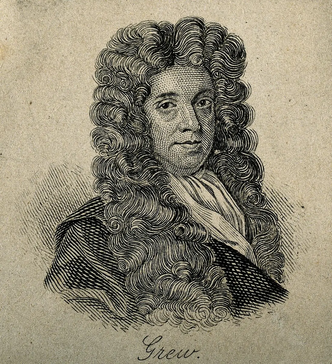
1.1.1 Concept of tissue
A single cell can do everything for itself only in the simplest living things. In a large plant or animal, cells divide the work: some cells carry water, some make food, some protect, some hold the body up. Cells that share a job usually share an origin and a shape too, and they lie together. Such a group is a tissue: a group of cells of common origin that carry out a particular function. Besides the cells, a tissue can contain non-living material between them, a matrix (in plants, mostly the cell walls and the material that cements them together).
Tissues are one level in the levels of biological organization:
Several tissues make an organ (a leaf, a root), organs make an organ system, and organ systems make an organism. Tissues are of two broad kinds: plant tissues and animal tissues. This unit is about plant tissues; animal tissues come in Unit 14.
Plant tissues
Plant anatomy is the study of the internal structure of plant parts, chiefly the structure and functions of their tissues. Tissues are too small to see with the eye, so the botanist cuts a very thin slice (a section) of a root, stem, leaf or flower bud and looks at it under a microscope. A cut straight across the organ is a transverse section (T.S.); one cut along its length is a longitudinal section (L.S.). A leaf is cut across from its top surface to its bottom one, a vertical section (V.S.).
Tissues build the plant’s organs, and organs work together as organ systems. A flowering plant has two organ systems, named by where they are:
- the shoot system, usually above ground. Its vegetative (non-reproductive) parts are the stem and leaves, which hold the plant up to the light and make food by photosynthesis; its reproductive parts are the flowers and the fruits with their seeds;
- the root system, usually below ground. It anchors the plant, takes in water and minerals, and often stores food (a radish, a carrot).
1.1.2 Types of plant tissues
There are two ways to sort a plant’s tissues, and both are used.
By where they are and what they do, a vascular plant (one with xylem and phloem) has three tissue systems:
- Epidermal tissue system: the epidermis, the outer layer of cells covering the leaves and the young stems and roots. It protects, and in roots it takes in water.
- Ground tissue system: everything between the epidermis and the vascular tissue (cortex, pith and the leaf’s mesophyll). Its cells are the least specialized; they make food by photosynthesis and store it.
- Vascular tissue system: the xylem and phloem, which carry materials through the plant and help to support it.
By whether their cells can still divide, plant tissues are of two kinds:
- a.meristematic tissue, cells that keep dividing;
- b.permanent tissue, cells that have stopped dividing and taken up a fixed job.
1. Meristematic tissue
A meristematic tissue, or meristem (Greek meristos, divided), is a group of actively dividing cells in the growing parts of a plant. Its cells are built for one job, dividing by mitosis:
- they are young and undifferentiated: not yet changed into any particular kind of cell;
- they are small, living, with thin cellulose walls;
- they are isodiametric (about as wide as they are long), oval, round or many-sided;
- they are packed closely, with no intercellular spaces;
- each has a large, conspicuous nucleus and dense cytoplasm;
- they have no stored food and no cell inclusions (crystals, tannins), and vacuoles are small or absent;
- their rate of metabolism is high, as they must build new cells all the time.
The meristems are what make a plant grow. An animal grows all over its body until it is adult and then stops; a plant keeps meristems at its tips and in its stems all its life, so a tree can grow for hundreds of years.
Encyclopaedia Britannica (1911), "Plants", public domain, via Wikimedia Commons
Root tip: New Student's Reference Work (1914), public domain
Types of meristem
Meristems are sorted in three ways: by their origin and the growth they make, by their position in the plant, and by the planes in which their cells divide.
A. By origin and growth
Before these two, there is the promeristem (primordial meristem): the very first cells of a growing tip, in the embryo and at the extreme tip of the root and shoot, from which all other meristems come.
- a.Primary meristem: it comes straight from the embryo’s meristem and has never stopped dividing. It makes the primary growth of the plant: the plant grows longer, its roots go deeper, and new leaves and branches form. Every plant shows primary growth. The apical and intercalary meristems, and the cambium inside a dicot’s vascular bundle, are primary.
- b.Secondary meristem: it forms later, from permanent cells that become able to divide again (they de-differentiate). It makes the secondary growth of the plant: the stem and root grow thicker. Secondary growth is found mainly in dicots and gymnosperms. Examples: the cork cambium and the interfascicular cambium (both met in 1.1.6).
B. By position
Seedling: V. Rattan, A Popular California Flora (1888), public domain
Sugar cane: P. J. F. Turpin (c. 1820), public domain
- a.Apical meristem: at the tips (apices) of the plant’s parts: the root tip, the shoot tip, the tips of branches and flower buds. It increases the length of the plant, so the shoot grows taller and the root deeper. Examples: the root apex, the shoot apex.
- b.Lateral meristem: along the sides of the stem and root, in a cylinder parallel to the surface (it looks like a ring in a cross section). It increases the thickness (girth, diameter) of the plant. Examples: the vascular cambium and the cork cambium of dicot stems and roots.
- c.Intercalary meristem: intercalary means “put in between”. It lies between permanent tissues, at the base of the internodes or of the leaves. It is a piece of the apical meristem that was left behind, separated from the tip as the stem grew. It lengthens the internodes, and helps a flattened (lodged) cereal or grass to stand up again: the cells on the lower side of the joint grow longer, and the stem bends upward. It is common in monocots: grasses, bamboo, wheat, maize, sugarcane, and the mint’s internodes too. It usually works for a short time and then becomes permanent tissue. It is absent in rosette plants such as cabbage and cauliflower, whose internodes stay very short, so their leaves are crowded together.
C. By the planes of division
- a.Rib (file) meristem: its cells divide in one plane only, at right angles to the length of the row, so the row just gets longer: it builds long rows (files) of cells. In a young stem, rib meristem below the shoot tip builds the pith and cortex as columns of cells.
- b.Plate meristem: its cells divide in two planes, so a single layer spreads out sideways into a sheet. It increases the surface area of a flat organ, such as the leaf blade.
- c.Mass meristem: its cells divide in all planes, so it grows into a solid mass and increases the volume of the organ: the embryo, the endosperm, a young fruit or ovary, the corpus of a shoot tip.
Which meristem makes a sugarcane stem’s internodes longer?
Pick an answer to see why.
A leaf grows from a tiny bump into a broad, thin blade. Which kind of meristem makes it spread out that way?
Pick an answer to see why.
Functions of meristems
- The promeristem brings about the embryo’s growth and gives rise to all the other meristems; the primary meristems carry out primary growth and the secondary meristems secondary growth.
- The apical meristem increases the height of the plant and the length of its roots; the lateral meristem increases the girth (the cross-sectional area); the intercalary meristem increases the length of the internodes in maize, sugarcane and other monocots.
- Just behind the tip, the apical meristem’s new cells form three primary meristems: the protoderm forms the epidermis; the ground meristem forms the ground tissues (cortex, endodermis, pericycle, pith); the procambium forms the vascular tissues (xylem, phloem, and the cambium between them).
- An older way of describing the same layers, the histogen theory (Hanstein, 1868), names the layers of the root tip: the dermatogen gives rise to the epidermis; the periblem to the cortex and endodermis; the plerome to the stele (pericycle, vascular bundles, pith and medullary rays); and in a root, the calyptrogen to the root cap. In the shoot tip the layers are less clear, and the tunica–corpus theory (Schmidt, 1924) fits better: an outer tunica of one or two layers, whose cells divide only at right angles to the surface, round an inner corpus whose cells divide in all planes.
2. Permanent tissue
When a meristem’s cells stop dividing, they grow, take a fixed shape and size, and become specialized for a job: they differentiate. A group of such mature, differentiated cells is a permanent tissue. Its cells:
- may be alike or of several kinds;
- may be living or dead (some cells do their job best as empty, dead walls), or a mixture;
- have lost the power of division, so permanent tissues don’t make the plant grow;
- do other jobs: storing food, support, protection, transport.
By the kinds of cell they contain, permanent tissues are of three groups:
- a.simple permanent tissue: one kind of cell;
- b.complex permanent tissue: several kinds of cell working together;
- c.special (secretory) tissue: cells or groups of cells that make and release substances.
a. Simple permanent tissue
A simple permanent tissue is made of one kind of cell (it is homogeneous), all of common origin and alike in shape, size and function. There are three: parenchyma, collenchyma and sclerenchyma.
i. Parenchyma
Parenchyma (Greek para, beside, enchyma, something poured in) is the most common tissue in plants and the least specialized. Its cells are:
- living, each with a nucleus, cytoplasm and usually a large central vacuole;
- oval, round or many-sided, about as long as they are wide;
- thin-walled, the walls of cellulose;
- loosely packed, with intercellular spaces where three or more cells meet.
Parenchyma fills the soft parts of plants: the cortex and pith of stems and roots, the flesh of fruits, the mesophyll of leaves; there is parenchyma in the xylem and phloem too. Its main job is to store food (and water). Because its cells are alive and little specialized, parenchyma can also take on other jobs, and even start dividing again to heal a wound. Its special kinds:
- Epidermis (in the root called epiblema): a single layer of parenchyma cells forming the outer surface of the root, stem and leaves, with no intercellular spaces. It protects the parts inside, and in roots it takes in water and minerals.
- Chlorenchyma: parenchyma whose cells contain chloroplasts. It makes food by photosynthesis. It forms the mesophyll of leaves and the outer cortex of young green stems.
- Aerenchyma: parenchyma with large air spaces (air cavities) between its cells. It is common in water plants, such as the lotus, water hyacinth and Hydrilla: the air makes the plant light and buoyant, so it floats, and lets oxygen reach the parts under water.
- Prosenchyma: parenchyma whose cells are long, with tapering ends, thickish walls and close packing, but still living. It gives extra support, in the pericycle and the conjunctive tissue, for example.
- Idioblasts: single large parenchyma cells, scattered among ordinary ones, that store special substances: tannins, oils, crystals (of calcium oxalate, for example), mucilage. (Idios is Greek for “one’s own”: a cell different from its neighbors.)
Encyclopaedia Britannica (1911), "Plants", public domain, via Wikimedia Commons
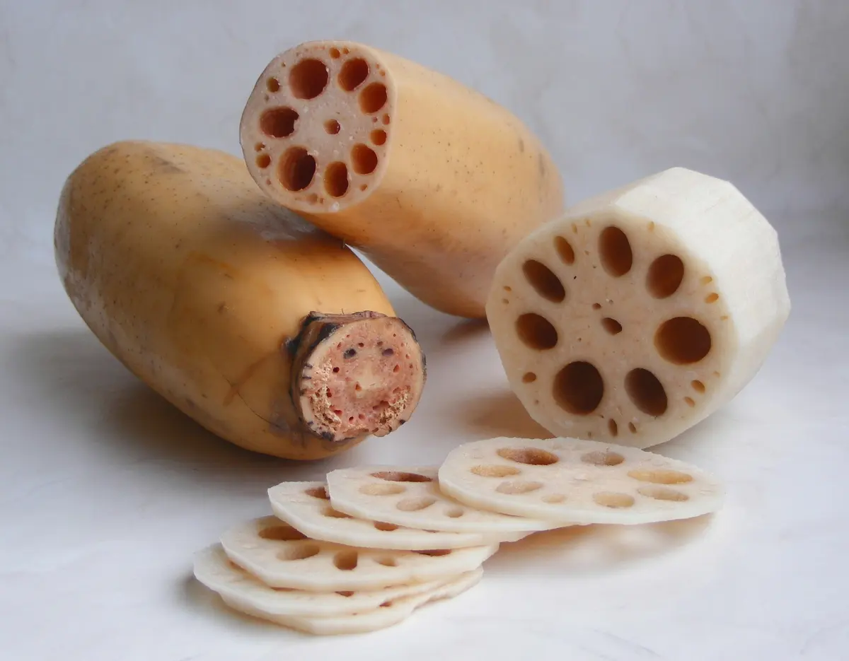
Which tissue makes the leaves and stems of a water hyacinth float?
Pick an answer to see why.
ii. Collenchyma
Collenchyma (Greek kolla, glue) is made of living, elongated cells (oval or many-sided in a cross section) whose walls are thickened unevenly: the thickening is laid down in some places only, mostly at the corners. The thickening is of cellulose, hemicellulose and pectin, and it holds a lot of water, which makes the walls strong but flexible. The cells are packed closely, with little or no intercellular space. Like parenchyma, each cell has a nucleus, cytoplasm and a vacuole, and sometimes chloroplasts.
Collenchyma lies just under the epidermis (as the hypodermis) in the young stems of dicots, in leaf stalks (petioles) and flower stalks (pedicels), and along the midrib of dicot leaves. It is usually absent in monocots, whose supporting tissue is sclerenchyma, and absent in roots.
It is called a living mechanical tissue, because it both supports the plant and stays alive. By how its walls are thickened, there are three kinds:
- Angular collenchyma: the walls are thickest at the corners where several cells meet (Datura, tomato, the stems of many herbs).
- Lacunar collenchyma: the thickening is on the walls next to intercellular spaces (lacunae) (Salvia, Cucurbita).
- Lamellar (plate) collenchyma: the thickening is on the tangential walls, the inner and outer walls of each cell, so it looks like layers or plates (sunflower’s hypodermis).
Snowman frosty, public domain, via Wikimedia Commons
Functions of collenchyma:
- It gives flexibility to plant parts and lets them bend without breaking: tendrils, the soft stems of climbers, leaf stalks in the wind.
- It keeps the soft stems of herbs from falling over (lodging) and lets young stems grow upright.
- It keeps leaves from tearing in the wind (at the midrib and edges).
- It gives mechanical strength to young stems, petioles and leaves while they are still growing (a dead, stiff tissue would stop them lengthening).
- Where it has chloroplasts, it carries out photosynthesis, and it stores some food.
iii. Sclerenchyma
Sclerenchyma (Greek skleros, hard) is made of cells whose walls are very thick, hard and impermeable, because of lignin laid down in the cellulose wall. Lignin makes the wall waterproof, so the cell’s contents die: mature sclerenchyma cells are dead, an empty space (the lumen) inside a thick wall. The cells are packed closely, with no intercellular spaces. The wall is crossed by thin channels, pits, where the lignin was not laid down.
Sclerenchyma does no metabolic work (no photosynthesis, no food storage). Its job is strength, rigidity, hardness and protection. It is found in the hypodermis of monocot stems (maize, sugarcane, bamboo), as the bundle caps and bundle sheaths round vascular bundles, as the bast fibers of the phloem of jute, hemp and flax, in the hard seed coats of beans and guava’s seeds, and in the hard shells (endocarps) of coconut, mango, almond and walnut (ओखर, Juglans regia).
By the shape of its cells, sclerenchyma is of two kinds:
- Fibers: very long, narrow, spindle-shaped cells with tapering, pointed ends, overlapping one another in strands. A fiber can be over a hundred times as long as it is wide. Fibers are named by where they are:
- bast (phloem) fibers, in the phloem and outer stem: jute, flax (linen), hemp, Nepal’s लोक्ता (Daphne) and अल्लो (Himalayan nettle);
- wood (xylem) fibers, in the xylem;
- surface fibers, on the outside of seeds and fruits: the coir of a coconut’s husk. (Cotton is often listed here too, but cotton’s “fibers” are really long hairs of the seed’s skin, made of almost pure cellulose with no lignin, which is why cotton cloth is so soft.)
- Sclereids (stone cells): short cells, about as long as they are wide (isodiametric) or of odd shapes, with walls even more heavily lignified than fibers, so thick that the lumen is tiny and the wall is layered and crossed by branching pits. They give hardness and protection: the shells of nuts, the stones of fruits, the hard coats of seeds, and the gritty grains in a pear or a guava.
Snowman frosty, public domain, via Wikimedia Commons
Plant cells: Encyclopaedia Britannica (1911), "Plants", public domain
Functions of sclerenchyma:
- Fibers give mechanical support to plant parts, strength without much weight.
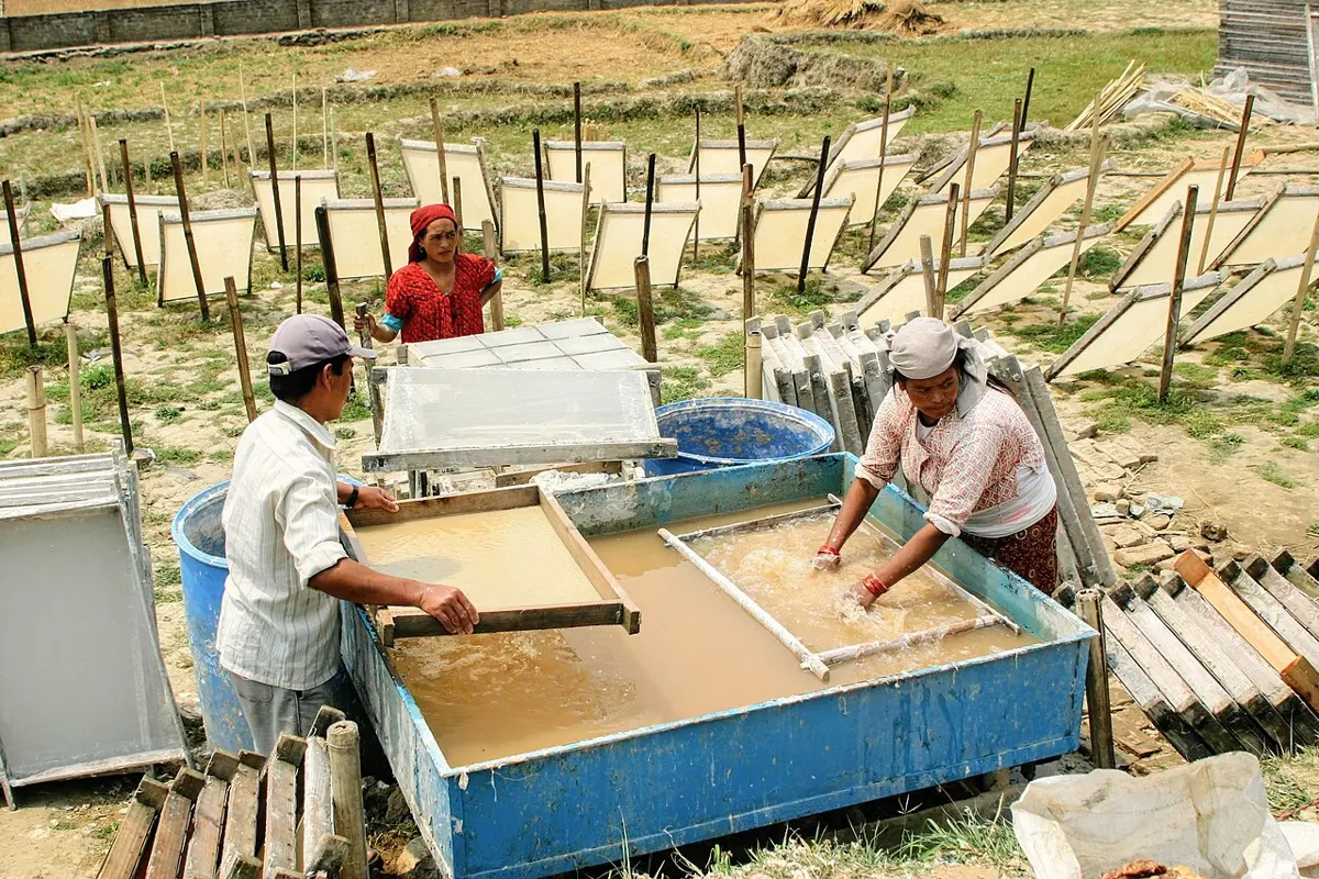
- The fibers of jute, hemp, flax and others are used to make sacks, rope, cloth and paper.
- Sclereids make plant parts hard and stiff.
- In almond, coconut and walnut, sclereids form the stony endocarp (the shell).
- In the seeds of legumes (beans, peas), sclereids form the hard protective seed coat (testa).
- The grittiness of guava, pear and some apples comes from clusters of sclereids in the soft flesh.

Which tissue makes a pear’s flesh feel gritty?
Pick an answer to see why.
| Parenchyma | Collenchyma | Sclerenchyma | |
|---|---|---|---|
| cells | living | living | dead when mature |
| shape | round, oval or many-sided | elongated | long fibers, or short sclereids |
| wall | thin, cellulose | thickened unevenly (cellulose, pectin) | thick all round, lignified |
| intercellular spaces | present | few or none | none |
| main job | storing food; photosynthesis | flexible support | rigid support, hardness |
| found in | soft parts of all organs | under the epidermis of dicot stems, petioles | monocot hypodermis, bundle caps, seed coats, shells |
b. Complex permanent tissue
A complex permanent tissue is made of several kinds of cell (it is heterogeneous), different in origin, shape, size and nature (some living, some dead), all working together for one function. There are two: xylem and phloem. Their work is conduction (transport), so they are called conducting tissues, and because they make up the vascular bundles (Latin vasculum, a little vessel), they are also called vascular tissues.
i. Xylem
Xylem (Greek xylon, wood) is also called wood or hadrome. It is made of four kinds of cells: tracheids, vessels, xylem parenchyma and xylem fibers. Tracheids and vessels, the cells that carry water, are together called the tracheary elements; in flowering plants, the vessels are the main ones.
Xylem carries water and dissolved minerals (the sap) in one direction, upward: from the roots, through the stem and branches, to the leaves, flowers and fruits. Most of its cells are dead and lignified, so it also gives the plant mechanical strength: the wood of a tree is xylem.
Plant cells: Encyclopaedia Britannica (1911), "Plants", public domain
Tracheids are elongated, dead cells with thick, lignified walls, a fairly wide empty lumen, and tapering, closed ends that overlap those of the next tracheid. Water passes from one tracheid to the next through pits in their walls. Tracheids are the main water-conducting cells in the xylem of pteridophytes and gymnosperms (a pine’s wood is almost all tracheids). In flowering plants they help the vessels. Their thick walls also make them strong.
Vessels are long tubes made of many short, wide, dead cells, the vessel elements (vessel members), placed end to end. The end walls between them, flat (transverse) or slightly slanting (oblique), dissolve completely or partly, leaving a hole, the perforation plate, so the row becomes one long, open channel. Water flows through a vessel much more easily than it seeps from tracheid to tracheid through pits. Vessels are linked to tracheids and to one another through pits in their side walls. They are the main conducting cells of the xylem of angiosperms, and are absent in most pteridophytes and gymnosperms (only a few, such as Gnetum and the bracken fern, have them). Their thick walls also give support.
As a tracheary element matures, a secondary wall of lignin is laid down inside its thin primary wall, in one of five patterns:
- Annular: separate rings;
- Spiral: a spiral band, like a spring;
- Scalariform: bars across the wall like the rungs of a ladder (Latin scala, ladder);
- Reticulate: a network of bands;
- Pitted: the whole wall thickened, except for small thin spots, the pits.
Rings and spirals cover little of the wall, so the cell can still stretch: they are found in the first xylem, which forms while the organ is still lengthening. Pitted walls are the strongest and are found in xylem formed after lengthening stops.
Xylem parenchyma: small, thin-walled, living cells among the xylem’s dead cells, many-sided in outline, with no intercellular spaces. They store food (starch, fats) and tannins, and carry sap slowly sideways (laterally). They are the only living cells of the xylem.
Xylem fibers (wood fibers): dead, long, thick-walled sclerenchyma fibers in the xylem. They give mechanical support and rigidity.
Which of these cells in the xylem is alive?
Pick an answer to see why.
Primary and secondary xylem
Primary xylem forms during primary growth, from the procambium. It forms in two stages:
- protoxylem (“first xylem”) forms first, while the organ is still growing in length; its vessels and tracheids are small (narrow) and have annular or spiral thickenings, which stretch;
- metaxylem (“later xylem”) forms after it, once lengthening is over, but before any secondary xylem; its vessels and tracheids are larger (wider), with reticulate or pitted thickenings.
Secondary xylem forms later, during secondary growth, from the vascular cambium. It makes up the wood of gymnosperms and dicot trees. Monocots have none, because they have no vascular cambium.
Position of protoxylem and metaxylem
Protoxylem and metaxylem are easy to tell apart in a section: small vessels against big ones. Where the small ones lie, relative to the big ones and to the center of the organ, gives four kinds of xylem:
- Endarch (endo, inside; arche, beginning): protoxylem towards the center (the pith), metaxylem towards the periphery (outside). The xylem grew from the inside outward. Found in stems (and the leaf bundles that continue from them).
- Exarch (exo, outside): protoxylem towards the periphery, metaxylem towards the center. The xylem grew from the outside inward. Found in roots.
- Mesarch (meso, middle): protoxylem in the middle, with metaxylem on both sides of it. Found in the stems of many ferns and in the leaves of cycads.
- Centrarch: protoxylem in the very center, surrounded on all sides by metaxylem. Found in the solid xylem core (protostele) of the earliest land plants, such as the fossil Rhynia.
ii. Phloem
Phloem (Greek phloios, bark) is also called bast or leptome. It is made of four kinds of cells: sieve tubes, companion cells, phloem parenchyma and phloem fibers. All are living except the phloem fibers.
Phloem carries food (sugars, mainly sucrose, and other organic substances) from where it is made or stored, the source (a leaf, or a potato in spring), to where it is used or stored, the sink (growing tips, roots, fruits, tubers). Sources and sinks change through the year, so the flow in phloem can go up or down: it is bidirectional, unlike the one-way flow in xylem.
Plant cells: Encyclopaedia Britannica (1911), "Plants", public domain
Sieve tubes are long tubes of wide, elongated, living cells, the sieve tube elements, placed end to end. Their end walls, flat or slanting, are full of pores, like a sieve: these sieve plates let the food pass from one element to the next through the sieve pores. A mature sieve tube element has no nucleus; it keeps a thin layer of cytoplasm round its wall and an open middle, so the sap flows freely. Sieve tubes are the main food-conducting cells.
Companion cells are narrow, elongated, living cells pressed against the side of each sieve tube element. A sieve tube element and its companion cell come from the same parent cell, which divided by mitosis, so they are sometimes called sister cells. A companion cell has a thin wall, a large active nucleus and dense cytoplasm, full of mitochondria. It is joined to its sieve element by many fine strands (plasmodesmata), and its nucleus controls the work of the sieve tube, which has none of its own: hence the name. Companion cells load sugar actively into the sieve tubes at the source and take it out at the sink, using energy.
In the phloem of pteridophytes and gymnosperms, there are no sieve tubes and no companion cells: the conducting cells are sieve cells, and their nuclear work is done by special parenchyma cells, the albuminous cells.
Phloem parenchyma: thin-walled, living, closely packed cells. They store food and other substances, and carry food slowly sideways. They are absent in the phloem of most monocots.
Phloem fibers (bast fibers): thick-walled, dead, elongated sclerenchyma fibers. They give mechanical support, and they are the fibers of jute, flax and hemp.
A sieve tube element has no nucleus. How does it stay alive?
Pick an answer to see why.
| Xylem | Phloem | |
|---|---|---|
| carries | water and minerals (sap) | food (sugars) |
| direction | one way, upward | both ways (source to sink) |
| main conducting cells | vessels and tracheids, dead | sieve tubes, living |
| cells | tracheids, vessels, xylem parenchyma, xylem fibers | sieve tubes, companion cells, phloem parenchyma, phloem fibers |
| living cells | only xylem parenchyma | all except phloem fibers |
| walls | thick, lignified | mostly thin, of cellulose |
| support | gives much support (wood) | gives little, through its fibers |
Vascular bundle
A vascular bundle is a strand of the plant’s vascular system: xylem and phloem, with or without a cambium between them. Bundles are seen in a section of any plant organ during its primary growth. They are sorted by how the xylem and phloem lie.
a. Radial bundles: the xylem and phloem are in separate strands, side by side on different radii (they alternate round a circle). Found in roots.
b. Conjoint bundles: xylem and phloem are joined into one bundle, on the same radius. Found in stems and leaves. There are three kinds:
- Collateral: the phloem lies on the outer side of the xylem, the two side by side on one radius.
- Open: a strip of cambium lies between them, so the bundle can make more xylem and phloem: secondary growth is possible. Example: the stem of sunflower and of most dicots and gymnosperms.
- Closed: no cambium, so no secondary growth. Example: the stems and leaves of monocots (maize, wheat, bamboo); the leaves of dicots.
- Bicollateral: the xylem lies between two patches of phloem, an outer and an inner one, with a cambium between each phloem and the xylem (outer cambium and inner cambium). Example: the stems of the cucumber family (cucumber, pumpkin, Cucurbita).
- Concentric: one tissue completely surrounds the other, rings round a core. Two kinds:
- Amphicribal (hadrocentric): xylem in the center, surrounded by phloem. Found in ferns (pteridophytes).
- Amphivasal (leptocentric): phloem in the center, surrounded by xylem. Found in the stems of Dracaena and Yucca (in the bundles made by their unusual secondary growth).
A bundle has phloem outside the xylem on one radius, and no cambium. What is it, and where would you find it?
Pick an answer to see why.
c. Special (secretory) tissue
Special tissues, or secretory tissues, are formed from simple or complex tissues that have become specialized to make (secrete) or store substances, or to get rid of waste (excrete). Many have a cavity or canal where the substance is stored until it is released. There are two kinds.
i. Laticiferous tissue
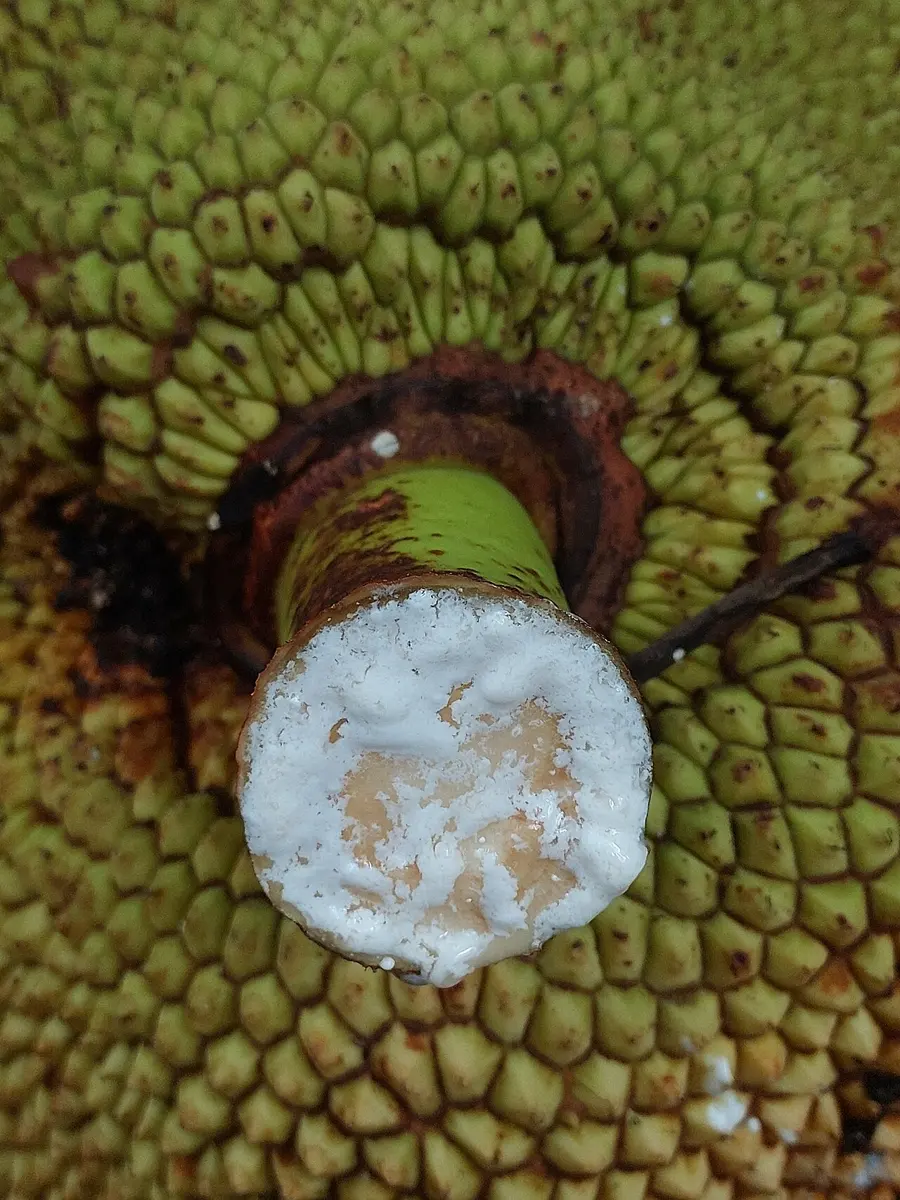
Laticiferous tissue makes and holds latex, the milky, white or yellow juice that oozes from a broken stem or leaf. Latex is a mixture: sugars, proteins, gums, alkaloids, rubber, and enzymes such as proteases (which break down proteins) and chitinases (which break down chitin, the material of insects’ skeletons and of fungal walls). It is sticky and often poisonous, so it defends the plant against insects, grazing animals and microbes, and it seals wounds, clotting over a cut like blood. Laticiferous tissue is of two kinds:
- latex cells (non-articulated laticifers): single cells that grow and branch through the plant, sometimes for meters, without dividing: in Euphorbia (सिउँडी), the oleander, and the fig family (Ficus: banyan, peepal, rubber plant);
- latex vessels (articulated laticifers): rows of cells whose end walls dissolve to make long tubes, often joined in a network: in papaya, banana, the rubber tree (Hevea), the opium poppy, and lettuce.
Other plants with latex include mulberry (Morus), jackfruit, the milkweed आँक (Calotropis) and hemp (Cannabis).
ii. Glandular tissue
Glandular tissue is made of glands: single cells or small groups of cells that make and release a particular substance, with or without a central cavity. Examples:
- oil glands in the peel of oranges and lemons (squeeze a peel near a flame and the oil sparks), and in the leaves of eucalyptus and tulsi;
- nectar glands (nectaries), which make sweet nectar for pollinating insects and birds;
- digestive glands on the leaves of insect-eating plants, such as the sundew (Drosera) of Nepal’s hills, which pour out enzymes to digest the insects they trap;
- resin ducts of pines, whose resin is tapped in Nepal’s chir pine forests for turpentine and rosin;
- stinging hairs of the nettle (सिस्नो): each is a single long cell with a brittle tip that snaps off in the skin and injects a mix of chemicals, including histamine;
- hydathodes, which let out drops of water at the tips of grass blades on a damp morning.
1.1.3 Anatomy of dicot and monocot root
In every section below, the tissues are described from the outside in, the way you would meet them looking from the edge of the slide towards its center. Learn the order of the layers first; the details then hang on it.
The central part of a root or stem, inside the endodermis, is the stele (Greek, a pillar): the pericycle, the vascular tissue and the pith. Everything outside it, from the epidermis to the endodermis, is the extra-stelar region.
A. Anatomy of dicot root
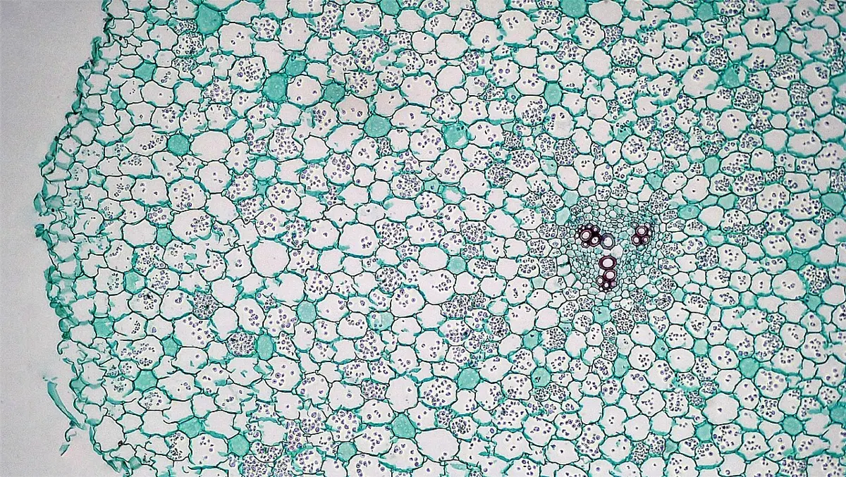
A dicot root (gram, bean, mustard, Ranunculus) cut across while it is young shows these tissues:
- Epiblema (piliferous layer, rhizodermis): the outermost single layer of thin-walled, closely packed parenchyma cells. It has no cuticle and no stomata (a waterproof cuticle would stop the root taking in water). Some of its cells grow out as long, unicellular root hairs, which take in water and minerals from the soil. It also protects the tissues inside.
- Cortex: a wide zone of many layers of thin-walled, round or oval parenchyma cells with intercellular spaces. It has no chloroplasts (no light reaches it). It stores food (starch) and passes water and minerals inward from the epiblema.
- Endodermis: the innermost layer of the cortex, a single ring of barrel-shaped living cells with no spaces between them. Its cells carry a band of suberin (a waxy substance) and lignin round their radial and transverse walls, the Casparian strip, which water can’t pass. So water and minerals can’t slip between the cells (through their walls) into the stele: they must go through the cells’ living membranes, which choose what to let in. As the root ages, most endodermal cells add more suberin and thicker walls; a few, opposite the arms of xylem, keep only their Casparian strips and stay thin-walled: these passage cells let water pass easily from the cortex to the xylem. Because it checks everything going into the stele, the endodermis is called the root’s biological check post.
- Pericycle: just inside the endodermis, one layer (sometimes a few) of thin-walled parenchyma cells with no intercellular spaces. It is the outer layer of the stele. Its cells keep the power to divide: lateral roots grow from it, and in secondary growth it forms part of the vascular cambium and the cork cambium.
- Vascular bundles: radial: xylem and phloem in separate strands, alternating on different radii. There are two to six xylem strands (diarch to hexarch; four, tetrarch, is common), with as many phloem strands.
a. Xylem: long strands radiating from the center, like the arms of a star, made of vessels, tracheids and xylem parenchyma (fibers are usually absent in a young root). The vessels are many-sided (polygonal) or angular, in a row along each arm. The xylem is exarch: protoxylem at the outer tip of each arm, against the pericycle, metaxylem towards the center, where the arms often meet. It carries water and minerals up, and gives support.
b. Phloem: small half-moon-shaped patches between the xylem arms, made of sieve tubes, companion cells, phloem parenchyma and a few fibers. It carries food. - Conjunctive tissue: parenchyma (thin-walled, closely packed, somewhat elongated cells) between the xylem and phloem strands. It stores food, and part of it becomes the vascular cambium when secondary growth begins.
- Pith (medulla): the central parenchyma. In a dicot root it is very small or absent, because the metaxylem fills the center. Its cells, round to many-sided, store food.
B. Anatomy of monocot root
A monocot root (maize, wheat, onion, orchids) cut across shows the same layers in the same order, with a few telling differences.
- Epiblema: a single layer of closely packed, thin-walled parenchyma cells with no intercellular spaces, no cuticle and no stomata, bearing unicellular root hairs.
- Cortex: a wide zone of many layers of round or oval parenchyma cells with intercellular spaces, which hold air for respiration and store starch; it passes water inward. In older roots the outer layers of the cortex become thick-walled (suberized or lignified) to form an exodermis, a protective layer once the epiblema and its hairs have worn away.
- Endodermis: the innermost layer of the cortex, a definite ring of barrel-shaped cells round the stele, with Casparian strips on their radial and transverse walls. In older roots, most endodermal cells also thicken their inner and side walls heavily (they look U-shaped in section); the few left thin-walled, opposite the protoxylem, are the passage cells. The endodermis controls what moves into and out of the stele, and stops air getting into the xylem’s vessels.
- Pericycle: usually one layer of thin-walled parenchyma just inside the endodermis (several layers in Smilax, कुकुरडाइनो): the outermost layer of the stele. It gives rise to lateral roots only (there is no secondary growth). In older roots it may become sclerenchyma.
- Vascular bundles: radial, xylem and phloem alternating on different radii, and many of them: polyarch, usually more than six strands each (a maize root may have twenty or more).
a. Xylem: vessels, tracheids, xylem parenchyma and xylem fibers. Exarch: the small protoxylem vessels, with annular or spiral thickenings, lie outside, against the pericycle; the large metaxylem vessels, round in section, with reticulate or pitted thickenings, lie inside. It carries water and minerals up and gives support.
b. Phloem: patches between the xylem strands, of sieve tubes, companion cells and phloem parenchyma (fibers usually absent). It carries food. - Conjunctive tissue: parenchyma between the xylem and phloem strands; it may become sclerenchyma in older roots.
- Pith: large and well developed, in the middle of the root: thin-walled, round or oval parenchyma cells, with or without spaces, storing plenty of starch.
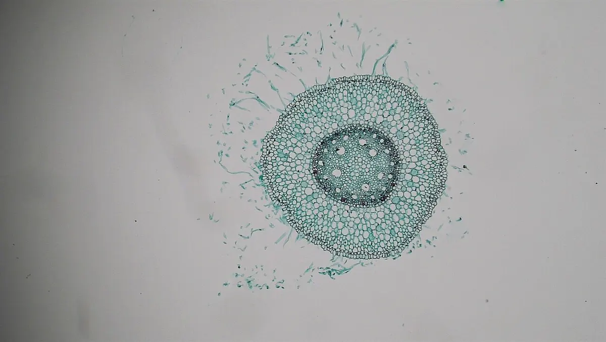
In a section of a root you see 14 xylem strands alternating with 14 phloem strands round a large pith. What is it?
Pick an answer to see why.
| Dicot root | Monocot root | |
|---|---|---|
| number of xylem strands | 2 to 6 (diarch to hexarch) | many, usually over 6 (polyarch) |
| xylem vessels | many-sided (angular) | round, the metaxylem large |
| pith | small or absent | large, well developed |
| conjunctive tissue | parenchyma; part becomes cambium | parenchyma, may become sclerenchyma |
| pericycle | gives rise to lateral roots, part of the cambium and the cork cambium | gives rise to lateral roots only |
| exodermis | absent | often present in older roots |
| secondary growth | takes place | does not take place |
1.1.4 Anatomy of dicot and monocot stem
A. Anatomy of dicot stem
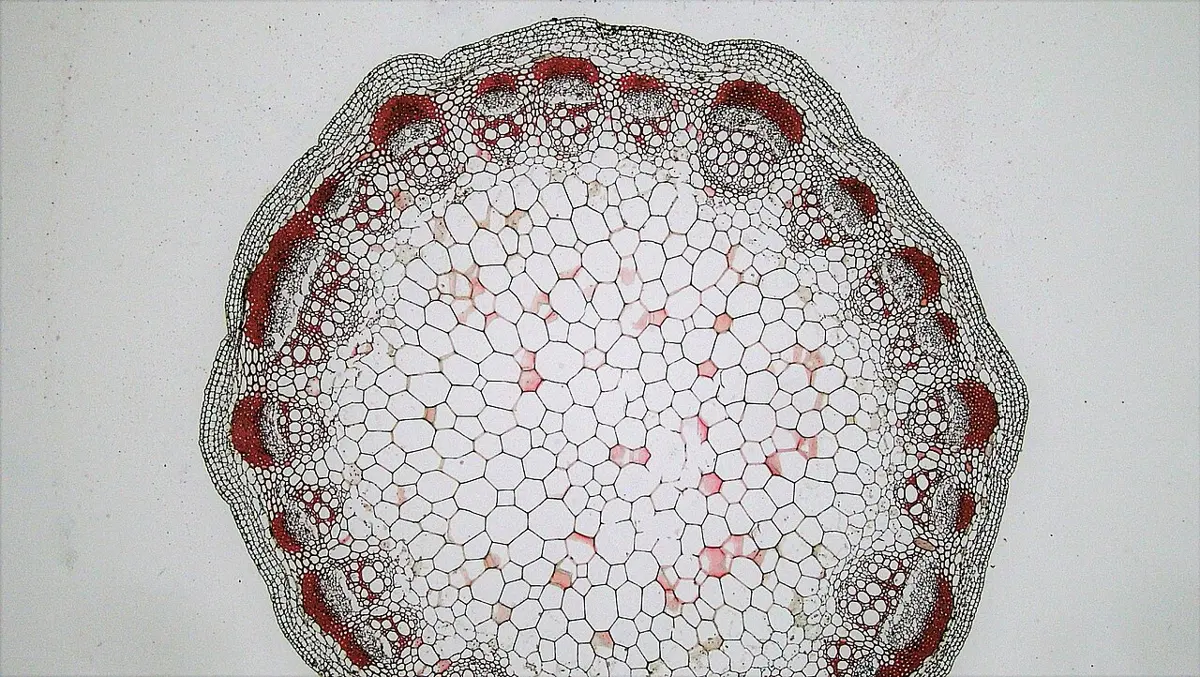
A young dicot stem (sunflower, Helianthus, is the usual example) cut across shows these tissues:
- Epidermis: the outermost single layer of closely packed, somewhat elongated parenchyma cells, transparent, with no chloroplasts (except in its guard cells). Its outer wall is covered by a waxy cuticle, which cuts water loss. It bears multicellular hairs (trichomes) and a few stomata, each a pore between two kidney-shaped guard cells, for the exchange of gases.
- Hypodermis: just below the epidermis, three or four layers of collenchyma (lamellar collenchyma in sunflower), with no intercellular spaces, their walls thickened with cellulose and pectin. It gives the young stem strength and flexibility. Under each stoma the hypodermis is missing or thin, so air can reach the green cortex.
- General cortex: a narrow zone of round or oval, thin-walled parenchyma with intercellular spaces. In a young stem the outer cortical cells have chloroplasts and make food; the cortex also stores food. In sunflower, the cortex has resin (oil) ducts: canals running along the stem, each lined by a layer of small glandular cells that secrete into it.
- Endodermis: the innermost layer of the cortex, one cell thick, wavy, of barrel-shaped cells with no spaces between them. Casparian strips are usually absent in a stem. Its cells are full of starch grains, so it is called the starch sheath.
- Pericycle: a zone a few layers thick inside the endodermis, made of two tissues (heterogeneous): sclerenchyma in half-moon patches, the bundle caps, just outside the phloem of each bundle, and parenchyma between them, outside the medullary rays.
- Vascular bundles: many, arranged in a ring (this arrangement of the stele is a eustele). Each is conjoint, collateral and open and wedge-shaped. (In the stems of Cucurbita, the pumpkin family, the bundles are bicollateral.)
a. Xylem: on the inner side of the bundle, towards the pith: vessels, tracheids, xylem parenchyma and xylem fibers. The xylem is endarch: the small protoxylem vessels point towards the pith, the large metaxylem vessels outward. The vessels are many-sided, in radial rows. It carries water and minerals up.
b. Phloem: on the outer side of the bundle, just under the bundle cap: sieve tubes, companion cells, phloem parenchyma and a few fibers. It carries food.
c. Cambium: a narrow strip of primary meristem cells, a few layers thick, between the phloem and the xylem. Because it lies inside a bundle (Latin fasciculus, a little bundle), it is the intrafascicular (fascicular) cambium; in secondary growth it becomes part of the cambium ring. - Medullary rays (pith rays): bands of parenchyma, their cells stretched out radially, between neighboring bundles, joining the pith to the cortex. Through pits, the ray cells are in close touch with the conducting cells of the xylem and phloem. They carry food and water sideways (radially), and let gases pass between the cortex and the pith.
- Pith (medulla): the large central region of round or many-sided parenchyma with intercellular spaces. It stores food. In some stems, such as Cucurbita, the middle of the pith breaks down as the stem grows, leaving a hollow, the pith cavity.
B. Anatomy of monocot stem
A monocot stem (maize, Zea mays, is the usual example) cut across looks quite different:
- Epidermis: the outermost single layer of parenchyma cells, covered with a thick cuticle. There are usually no hairs. In a young stem it has a few stomata, each between two dumbbell-shaped guard cells (in grasses such as maize). It protects.
- Hypodermis: two or three layers of sclerenchyma just under the epidermis. It gives mechanical strength: a maize stalk or a bamboo stays upright in the wind.
- Ground tissue: all the rest of the stem: round or oval, living, thin-walled parenchyma with clear intercellular spaces. It is not divided into cortex, endodermis, pericycle, pith and medullary rays: it is all one (homogeneous). It stores food. (In bamboo and wheat the middle of the ground tissue breaks down, so the internodes are hollow.)
- Vascular bundles: scattered through the ground tissue (this kind of stele is an atactostele, Greek ataktos, without order). They are conjoint, collateral and closed: there is no cambium. The bundles near the edge are small and crowded; those towards the middle are larger and fewer. Each bundle is oval, wrapped in a bundle sheath of sclerenchyma fibers, thickest above and below.
a. Phloem: on the outer side (towards the edge of the stem): sieve tubes and companion cells in a neat pattern, with no phloem parenchyma. It carries food.
b. Xylem: on the inner side, few but distinct, round vessels arranged in a V or Y shape: two large metaxylem vessels at the arms of the V, on the outer side, and one or two smaller protoxylem vessels at its foot, towards the center: the xylem is endarch. As the stem lengthens, the oldest protoxylem is torn apart and the cells round it break down, leaving a space, the protoxylem lacuna (often called the water cavity), which holds water. A space made by cells breaking down is called lysigenous. The xylem carries water and minerals up and gives support.
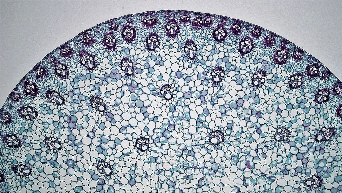
Which feature, on its own, shows that a stem section is a monocot’s?
Pick an answer to see why.
| Dicot stem | Monocot stem | |
|---|---|---|
| hairs | multicellular hairs present | usually none |
| hypodermis | collenchyma | sclerenchyma |
| ground tissue | divided into cortex, endodermis, pericycle, medullary rays, pith | one undivided ground tissue |
| bundles | in a ring (eustele) | scattered (atactostele) |
| size of bundles | about equal | small at the edge, large in the middle |
| type of bundle | conjoint, collateral, open | conjoint, collateral, closed |
| bundle sheath | absent (bundle caps instead) | a sclerenchyma sheath round each bundle |
| xylem | many vessels in radial rows | few vessels in a V or Y; a water cavity |
| phloem parenchyma | present | absent |
| pith and medullary rays | present | absent |
| secondary growth | takes place | does not take place |
1.1.5 Anatomy of dicot and monocot leaf
A leaf is cut vertically, from its upper surface to its lower one, across the midrib and some of the blade.
A. Anatomy of dicot leaf
A dicot leaf (mango, sunflower, peepal) is dorsiventral: its upper and lower sides are built differently, because the upper side faces the sun and the lower side is in shade.
Epidermis: two of them.
- The upper epidermis (on the adaxial surface, the side towards the stem) is a single layer of closely packed, barrel-shaped parenchyma cells with a thick cuticle. It has few stomata or none.
- The lower epidermis (on the abaxial surface) has a thinner cuticle and many stomata, each between two kidney-shaped guard cells.
The cuticle checks water loss by transpiration; the stomata let gases in and out between the leaf’s inside and the air. (Botanists call the upper surface ventral and the lower dorsal, which is why such a leaf is dorsi-ventral.)
Mesophyll (meso, middle; phyllon, leaf): the chlorenchyma between the two epidermis layers, full of chloroplasts, the main site of photosynthesis. In a dicot leaf it is divided into two:
- a.Palisade layer: just under the upper epidermis, one or two layers of long, column-shaped cells standing side by side, closely packed (like the stakes of a fence, a palisade), with many chloroplasts. It catches most of the light.
- b.Spongy layer: above the lower epidermis: loosely packed, irregular, round or oval cells with fewer chloroplasts and large intercellular spaces full of air (air cavities). It is where gases move about inside the leaf. The large air space just inside each stoma is the substomatal cavity: water vapor collects in it and diffuses out through the stoma in transpiration.
Vascular bundles: many, of different sizes, in the midrib and veins, where they take the place of the mesophyll. The bundle of the midrib is the largest; they get smaller as the veins branch out towards the leaf’s edges. Each is conjoint, collateral and closed, wrapped in a bundle sheath of closely packed parenchyma.
- a.Xylem: towards the upper epidermis: vessels, tracheids, xylem parenchyma and fibers. It brings water and minerals into the leaf, and helps to support it.
- b.Phloem: towards the lower epidermis: sieve tubes, companion cells, phloem parenchyma and a few fibers. It carries the food made in the leaf to the rest of the plant.
Bundle sheath extensions: in the midrib, there is no mesophyll. The bundle lies in the middle, and above and below it, from the bundle sheath to the epidermis, are masses of collenchyma. Along the smaller veins, columns of parenchyma or sclerenchyma reach from the sheath to the epidermis. These bundle sheath extensions support the veins and protect them when the leaf is bent and twisted by wind and storms.
B. Anatomy of monocot leaf
A monocot leaf (maize, wheat, grass) usually stands up more or less vertically, both its faces lit about equally, and it is isobilateral: the two sides are built alike.
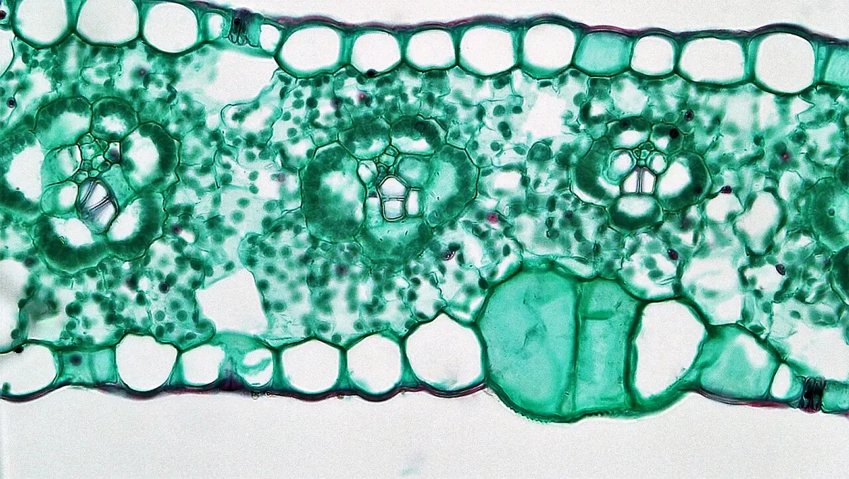
- Epidermis: an upper and a lower one, each a single layer of closely packed, barrel-shaped cells with a cuticle. Stomata are found on both surfaces, in about equal numbers, each between two dumbbell-shaped guard cells (in grasses). The epidermis protects, checks water loss and lets gases in and out through the stomata. In grasses (maize, rice, sugarcane), groups of large, thin-walled cells full of water, the bulliform cells (motor cells), lie in the upper epidermis. In hot, dry weather they lose water and shrink, so the leaf rolls up, shading its upper surface and cutting transpiration; when water is plenty, they swell and the leaf opens again.
- Mesophyll: undifferentiated: no palisade and spongy layers, just one kind of cell (homogeneous), round or oval, thin-walled, with air-filled spaces between them, containing chloroplasts. It carries out photosynthesis.
- Vascular bundles: many, parallel to one another (the parallel veins), of different sizes, large and small in turn. Each is conjoint, collateral and closed, surrounded by a bundle sheath of one layer of parenchyma (in maize, its cells hold large chloroplasts: a sign of the special C₄ photosynthesis you will meet in Unit 3).
a. Xylem: towards the upper epidermis: vessels, tracheids and xylem parenchyma; fibers are few. The vessels are round and lie in a V: the two large metaxylem vessels towards the lower epidermis (the phloem side), the small protoxylem towards the upper epidermis. Water and minerals travel in it, and it supports the leaf.
b. Phloem: towards the lower epidermis: sieve tubes, companion cells and a few fibers, no phloem parenchyma. It carries food out of the leaf. - Bundle sheath extensions: above and below the larger bundles (and in the midrib), the mesophyll is replaced by strands of sclerenchyma, reaching from the bundle sheath to the epidermis like girders. These dead, strong cells protect the bundles from damage, and keep the long thin leaf from tearing.
| Dicot leaf | Monocot leaf | |
|---|---|---|
| sides | dorsiventral: upper and lower sides differ | isobilateral: both sides alike |
| stomata | mostly or only on the lower surface | about equally on both surfaces |
| guard cells | kidney-shaped | dumbbell-shaped (grasses) |
| bulliform cells | absent | present in the upper epidermis of grasses |
| mesophyll | palisade and spongy layers | all one kind |
| veins (bundles) | a net of veins of many sizes | parallel bundles, large and small |
| bundle sheath extension | collenchyma (at the midrib) | sclerenchyma |
A leaf has palisade cells under its upper surface and almost all its stomata on its lower surface. What kind of leaf is it?
Pick an answer to see why.
1.1.6 Secondary growth of dicot stem
Primary growth, from the apical and intercalary meristems, happens in all plants: the shoot grows taller, the roots go deeper, and new leaves and branches form. Secondary growth is a second kind of growth, which makes the stem and root thicker: it increases their girth, diameter and cross-sectional area. It is found mainly in dicots and gymnosperms, and is absent in most pteridophytes and monocots, and in leaves.
Secondary growth is the work of the two lateral meristems, which make secondary tissues:
- the vascular cambium, which makes secondary xylem and phloem, inside the stele (intrastelar growth);
- the cork cambium, which makes the bark, outside it (extrastelar growth).
1. Activity of the vascular cambium
The vascular cambium adds new tissues inside the stele, so the stele widens. Its main product is secondary xylem, the wood, made up of heartwood and sapwood; each year it adds one more annual ring. It works in these steps.
a. Formation of the cambium ring. In a young dicot stem, each vascular bundle already has a strip of cambium between its xylem and phloem: the intrafascicular cambium (a primary meristem, left over from the procambium). When secondary growth begins, some cells of the medullary rays, in line with these strips, de-differentiate: they become meristematic again and start to divide. They form new strips of cambium between the bundles, the interfascicular cambium (a secondary meristem). The intrafascicular and interfascicular strips join up into a complete circle, the cambium ring.
The cambium ring is a single layer of cells (with the young cells it has just made on either side, it looks like a band a few cells thick). It has two kinds of cell, called initials:
- fusiform initials: long, spindle-shaped cells with tapering ends, lying lengthwise in the stem. They make the xylem and phloem.
- ray initials: short, almost isodiametric cells in small groups. They make the rays.
b. Formation of secondary vascular tissues. Each fusiform initial divides lengthwise (parallel to the stem’s surface) into two cells: one stays a cambium cell, the other becomes a tissue cell. A new cell cut off on the inner side becomes secondary xylem; one cut off on the outer side becomes secondary phloem. The cambium cuts off many more cells towards the inside than the outside, so most of what it makes is xylem, and the cambium itself is pushed outward as the wood piles up inside it.
As secondary xylem and phloem are laid down between them, the primary xylem and primary phloem, which were close together in each bundle, are pushed far apart: the primary xylem stays near the pith, in the very middle of the trunk; the primary phloem is pushed outward and, as the stem keeps widening, crushed. New layers of secondary xylem and phloem are added on the inner and outer sides of the cambium every growing season, as long as the plant lives.
c. Formation of secondary medullary rays. The ray initials divide to make narrow bands of living parenchyma cells, two or three cells wide, running radially (from the center outward) through the secondary xylem and the secondary phloem: the secondary medullary rays (vascular rays). They carry food inward from the phloem and water and minerals outward from the xylem, and store food. On a cut log they show as fine lines radiating from the center.
d. Formation of annual rings. The cambium’s work changes with the seasons. Where there is a cold or a dry season, it is very active in spring (or when the rains start): it makes many wide vessels with thin walls, to carry lots of water to the new leaves. This pale, open wood is the spring wood (early wood). Later in the year, in the cool or dry season, it slows down and makes fewer, narrower vessels and more fibers with thicker walls: the dense, dark autumn wood (late wood). Then it stops for the winter or the dry season.
One band of spring wood and the band of autumn wood after it make an annual ring (growth ring): the wood added in one year. Next spring the new, wide-celled spring wood starts right against the last year’s dense autumn wood, so the boundary between years is sharp, and the rings are easy to count.
So the age of a tree can be found, roughly, by counting its annual rings: this is dendrochronology (Greek dendron, tree; chronos, time). A forester need not cut the tree: a thin hollow drill, an increment borer, takes out a pencil-thin core from bark to center, and the rings are counted on it. Wide rings grow in good years, narrow ones in cold or dry years, so the pattern of ring widths is also a record of past climate: reading it is dendroclimatology.
Where the climate is the same all year, as in an evergreen rainforest that is warm and wet in every month, the cambium works at about the same rate all the time. There is no spring wood and autumn wood, and no distinct annual rings can be seen: the wood is uniform.
e. Formation of heartwood and sapwood. In an old tree, after many years of secondary growth, the wood near the center stops carrying water. Its living xylem parenchyma cells die, and before they die they fill the wood with tannins, resins, gums, oils and colored substances, which make it darker, harder, heavier and more durable: insects and fungi can’t easily attack it. Many of its vessels are also blocked by tyloses: balloon-like outgrowths of the neighboring parenchyma cells, which bulge into the vessel through its pits and plug it. This dark central wood is the heartwood (duramen). It no longer conducts; it only supports the tree.
The outer, younger wood, pale (often yellowish white), whose vessels and tracheids are still open (no tyloses) and whose parenchyma cells are still alive, is the sapwood (alburnum). It carries water and minerals and stores food. Each year the cambium adds a new ring of sapwood outside, and the innermost ring of sapwood turns into heartwood, so the heartwood keeps growing while the sapwood stays a band of about the same width.
Why is heartwood more durable than sapwood?
Pick an answer to see why.
2. Activity of the cork cambium
As the vascular cambium makes the stem thicker, the epidermis and the cortex outside it are stretched, and in time they tear. Before they do, some cells of the outer cortex (in some plants, of the epidermis or the hypodermis) become meristematic again and form another ring of cambium, outside the stele: the cork cambium, or phellogen. Its growth is extrastelar secondary growth, and it makes the periderm.
The cork cambium divides on both sides, but makes many more cells outward than inward:
- on the outer side, the phellem or cork: rows of dead, closely packed, box-shaped (rectangular) cells whose walls are thickened with suberin. Cork is waterproof and keeps out microbes; it protects the living tissues inside from infection, injury, heat, cold and water loss;
- on the inner side, a few layers of phelloderm or secondary cortex: living, thin-walled parenchyma cells, round or oval, with no intercellular spaces. It stores food.
These three layers together, phellem + phellogen + phelloderm, are the periderm. Everything outside the vascular cambium, the secondary phloem, the periderm and any crushed old tissues, is what we call bark.
Lenticels. Once the epidermis is torn and replaced by cork, its stomata are gone, and the cork is airtight. So at some places, usually where stomata used to be, the cork cambium makes loose, rounded, thin-walled cells with air spaces between them, the complementary cells, instead of cork. They push out through the bark as small raised pores, the lenticels, through which the living tissues inside exchange gases with the air (and lose a little water, lenticular transpiration). They show as the pale dots or short lines on a young branch, or the long horizontal bars on a birch.
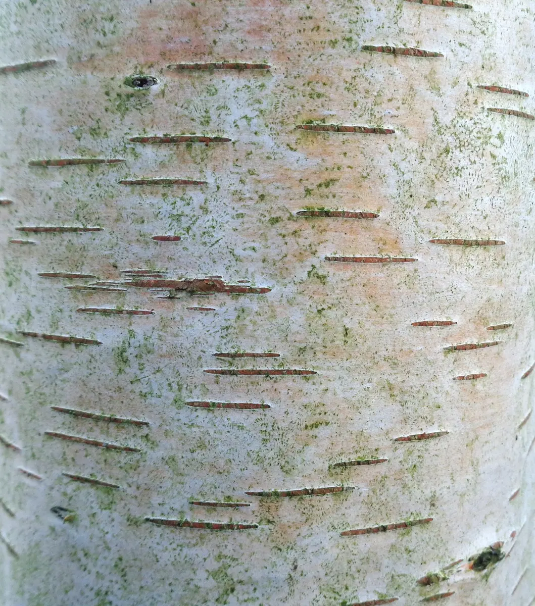
So secondary growth gives a dicot stem a central core of wood and an outer covering of periderm (bark).
Significance of secondary growth
- The wood made by the vascular cambium is commercially important: timber, furniture, paper, firewood.
- It makes the stem thicker, and its heartwood gives the mechanical support a tree needs to stand tall and upright for years.
- Counting the annual rings gives the approximate age of a tree.
- The rings of old trees record the climate of the past (dendroclimatology), which helps scientists understand how the climate is changing and what may come.
- The corky bark protects the inner tissues from injury, heat, cold and infection.
- Lenticels allow gas exchange (and some lenticular transpiration) through the bark.
- Each year it adds new xylem and phloem, replacing old vessels that have stopped working, and makes room for a growing tree’s ever larger crown of leaves.
Which tissues make up the periderm?
Pick an answer to see why.
Exercise
A. Choose the correct option
- Plant anatomy deals with:
Pick an answer to see why.
- Who is known as the father of plant anatomy?
Pick an answer to see why.
- Which tissue makes the outer lining of plant parts?
Pick an answer to see why.
- A meristematic cell has:
Pick an answer to see why.
- Which is the correct pair?
Pick an answer to see why.
- The lateral meristem increases:
Pick an answer to see why.
- Parenchyma that stores cell inclusions is:
Pick an answer to see why.
- The cell wall of collenchyma consists of:
Pick an answer to see why.
- Aerenchyma is commonly present in:
Pick an answer to see why.
- An elongated, narrow, non-living cell with tapering ends is:
Pick an answer to see why.
- The living mechanical tissue is:
Pick an answer to see why.
- The empty space inside a sclerenchyma fiber is called the:
Pick an answer to see why.
- Which of the following is a heterogeneous tissue?
Pick an answer to see why.
- The plant tissue that carries prepared food from the leaf to other parts is:
Pick an answer to see why.
- Which of the following is not a part of xylem?
Pick an answer to see why.
- All the cells of phloem are living except:
Pick an answer to see why.
- In the root of an angiosperm, the vascular bundles are:
Pick an answer to see why.
- When the xylem is surrounded by phloem on the outside, the vascular bundle is said to be:
Pick an answer to see why.
- In exarch xylem, the protoxylem lies:
Pick an answer to see why.
- Which of the following stops the conducting function of xylem elements?
Pick an answer to see why.
- Which is the correct pair?
Pick an answer to see why.
- In the leaf of a plant, which is true of the xylem?
Pick an answer to see why.
- As secondary growth goes on over the years, what happens in the trunk of a tree?
Pick an answer to see why.
B. Answer the following questions
Define meristem. Write its characteristic features.
Answer
Classify meristems by their position, and explain each with an example.
Answer
What is parenchyma? Discuss its modifications.
Answer
Compare a fiber with a sclereid.
Answer
Define complex permanent tissue and write its types.
Answer
Differentiate between xylem and phloem.
Answer
Study the figure above and answer:
- a.Name the type of vascular bundle.
- b.In which kind of plants are such bundles found in the stem?
- c.Is secondary growth possible in such a bundle? Why?
Answer
Mention the characteristic features of the anatomy of a dicot root.
Answer
Compare the anatomy of a dicot root and a monocot root.
Answer
Study the figure above and answer:
- a.Name the section.
- b.What is its hypodermis made of?
- c.Write about its vascular bundles.
Answer
Draw a labeled diagram of a monocot stem.
Answer
How does a dicot leaf differ from a monocot leaf? Answer on the basis of the distribution of stomata, the nature of the mesophyll, the shape of the guard cells and the bundle sheath extensions.
Answer
What is periderm? Explain its components with a labeled diagram.
Answer
Differentiate between early wood and late wood.
Answer
C. Long answer questions
Define plant tissue. Show the different types of plant tissues in a flow chart.
Answer
Describe the types of simple permanent tissues, with diagrams.
Answer
What are vascular bundles? Classify them by the arrangement of their tissues and describe each type with diagrams.
Answer
Discuss the anatomy of a monocot root with a suitable diagram. Mention any three diagnostic features.
Answer
What is secondary growth? Describe the growth of the stele, with relevant diagrams.
Answer
Printable PDFs
- Study notes 5 pages
- Worksheet 4 pages Answer key
Quiz yourself: Living Things and Their Structure, Grades 5–12 →
Practice every question in the book, and keep track of the ones you miss →
A companion to your textbook, not a replacement for it: study from the book your school uses, and where the two differ, follow your teacher. Found a mistake? Please tell us.