
A mosquito lands on your arm in the dark. Before you have even thought the word “mosquito”, your other hand has already slapped it. In that half second a message ran from a few touch receptors in your skin, up a nerve at about the speed of a motorbike on the highway, into your spinal cord and brain, and back out to the muscles of the other arm. The messages were not electricity in a wire. They were waves of sodium and potassium ions flicking in and out through tiny gates in the membranes of your nerve cells.
Your nervous system is about 86 billion such cells in the brain alone, linked by something like a hundred trillion junctions. It lets you see, think, speak, remember, keep your heart in time and your balance on a moving bus. This unit takes it apart: the brain and spinal cord and the layers that protect them, the nerves that leave them, the automatic system that runs your organs, and finally the nerve impulse itself, how it starts, how it travels, and how it crosses from one cell to the next.
Introduction
Your body has two systems that control and coordinate everything it does:
- the nervous system, which works by nerve impulses, electrical signals along nerve cells, fast (thousandths of a second) and aimed at particular muscles and glands;
- the endocrine system, which works by hormones, chemical messengers carried by the blood, slower (seconds to days) and reaching every cell that has a receptor for them.
The two work together as the neuro-endocrine system, which controls the body’s homeostasis (keeping its inside steady): metabolism, energy balance, the response to stress, growth and reproduction. Its center is the hypothalamus with the pituitary gland below it and their connections to the rest of the brain, together with other parts outside the brain that work like nerve tissue, such as the inner part of the adrenal glands, the adrenal medulla. You will meet the hormones in the next units; this unit is about the nerves.
Neurology is the branch of medicine that studies the nervous system and treats its disorders (a neurologist is its doctor).
All living things can sense a change around them and respond to it. This ability is called sensitivity, sensibility or irritability, and it is a property of protoplasm, the living matter of every cell. In animals with nerves, it happens in steps:
- Excitability: a receptor or nerve cell is excited by a stimulus;
- Reactivity: the neurons are ready to pass the signal on;
- Responsiveness: the signal causes a change inside the body;
- Behavior: the whole animal does something in answer.
All of these are carried out by neurons (nerve cells), which develop from the ectoderm of the embryo, the same layer that makes the skin. The neuron is the structural and functional unit of the nervous system.
Which of these is NOT a difference between the nervous system and the endocrine system?
Pick an answer to see why.
8.5.1 Types of nervous system
The nervous system is divided into parts by where they are and what they do:
- The central nervous system (CNS) is the brain and the spinal cord. It receives messages from the sense organs, decides what to do, and sends out orders.
- The peripheral nervous system (PNS) is all the nerves outside the CNS: 12 pairs of cranial nerves from the brain and 31 pairs of spinal nerves from the spinal cord. They connect the CNS with the receptors (in the eyes, ears, nose, tongue and skin) and the effectors (muscles and glands).
- The autonomic nervous system (ANS) is the part of the nervous system that runs the organs without our will: the heart, the gut, the glands, the blood vessels. It has two halves, the sympathetic and the parasympathetic.
Each message runs along three kinds of neuron:
- sensory (afferent) neurons carry impulses from receptors to the CNS;
- interneurons (association neurons) inside the CNS connect sensory and motor neurons and do the “thinking”;
- motor (efferent) neurons carry impulses from the CNS to effectors, the muscles and glands.
A sensory neuron carries an impulse
Pick an answer to see why.
1. Central nervous system: the brain
The meninges
The brain and spinal cord are soft, about as firm as tofu. They sit in bone, the cranium of the skull and the neural canal of the backbone, and between the bone and the nervous tissue lie three wrappings of connective tissue, the meninges (singular meninx, Greek for membrane). From outside in:
- Dura mater (Latin, “tough mother”): the outermost, thick, tough and fibrous. Around the brain it has two layers and is fixed to the inside of the skull; in places its two layers separate to enclose the big veins that drain the brain, the dural venous sinuses. Around the spinal cord it is a single tube. It carries its own blood vessels.
- Arachnoid mater (Greek, “like a spider’s web”): the middle layer, thin and delicate, without blood vessels. Fine web-like threads cross from it to the layer below.
- Pia mater (Latin, “gentle mother”): the innermost, very thin, transparent and rich in blood vessels, stuck closely to the surface of the brain and cord, following every fold.
Between them are spaces:
- the epidural space, between the bone and the dura. In the skull there is normally no real space, because the dura is fixed to the bone; in the backbone there is a real space, filled with fat and veins (it is where a doctor injects an epidural anesthetic for childbirth).
- the subdural space, between the dura and the arachnoid, normally only a thin film of fluid;
- the subarachnoid space, between the arachnoid and the pia, filled with cerebrospinal fluid (CSF). This is where the CSF is: the brain floats in it.
The CSF is made by the choroid plexuses, tufts of pia mater and capillaries that hang into the brain’s ventricles (below).
Servier Medical Art (smart.servier.com), CC BY 4.0
Which meninx is in direct contact with the brain and follows every fold of it?
Pick an answer to see why.
I. Brain
The brain is the main organ of the nervous system, the front part of the CNS, safe inside the cranium.
- It is about 98% of the weight of the CNS, and about 2% of the body’s weight: about 1.4 kg in a 70 kg adult.
- It grows fast in childhood: by the age of about six it has reached about 90% of its adult size.
- It receives about 15% of the blood the heart pumps and uses about 20% of the oxygen you breathe in, though it is only 2% of your weight. Most of that energy goes into pumping ions, to keep its neurons ready to fire (you will see why in 8.5.2).
- It holds about 86 billion neurons and about as many other cells, mostly neuroglia (glial cells), which support, nourish, insulate and protect the neurons. Glia make up about half the mass of the brain and spinal cord. (These counts come from a careful count of whole human brains published in 2009; the old claim of “100 billion neurons and ten times as many glia” was a guess.)
The brain’s tissue is of two kinds:
- gray matter, made mostly of the cell bodies of neurons (with their dendrites and the junctions between them). It looks gray because cell bodies have no fatty covering.
- white matter, made mostly of myelinated axons, nerve fibers wrapped in fatty myelin, which is white. These are the cables that join one area to another.
In the cerebrum and cerebellum, the gray matter is on the outside (the cortex) and the white matter inside. In the spinal cord it is the other way round: gray inside, white outside.
The brain develops from three swellings at the front of the embryo’s neural tube, and is divided into three parts: the forebrain, the midbrain and the hindbrain.
Patrick J. Lynch, medical illustrator, CC BY 2.5, via Wikimedia Commons
Turn the brain and spinal cord round, and tap a part to see its name. Notice how the cerebrum covers nearly everything, and how the cerebellum tucks under its back.
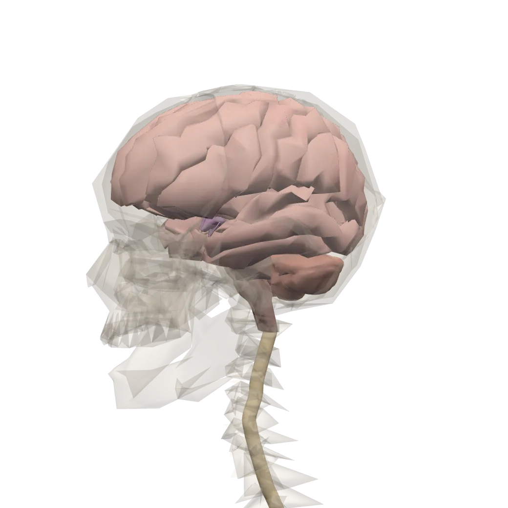
A model of a real body. Tap Show in 3D to turn it and tap its parts.
CerebrumThe largest part of the brain, in a left and a right hemisphere. It makes sense of what we see, hear, smell, taste and touch, and is the seat of thought, memory and speech. It controls the movements we choose to make.
CerebellumLies at the back, below the cerebrum. It keeps the body balanced and makes our movements smooth and well timed, as we walk, run or ride a bicycle.
Thalamus and hypothalamusDeep in the middle of the brain, above the brainstem. Most messages from the senses pass through the thalamus on their way to the cerebrum. The hypothalamus controls the pituitary gland, and through it several other glands.
MidbrainThe top part of the brainstem, which joins the brain to the spinal cord. With the pons it acts as a bridge, passing messages between the spinal cord and the higher parts of the brain.
PonsThe middle part of the brainstem, the rounded swelling in front of the cerebellum. Like the midbrain, it is a bridge for messages between the spinal cord and the higher parts of the brain.
Medulla oblongataThe lowest part of the brain, going on as the spinal cord. It runs the involuntary actions that keep us alive: breathing, the heartbeat and blood pressure, and also swallowing, coughing, sneezing and vomiting.
Spinal cordA long, soft cord inside the backbone, from the medulla oblongata down to about the first or second lumbar vertebra. It carries messages between the brain and the body, and is the center for most reflex actions.
Body and organs: BodyParts3D, The Database Center for Life Science (CC BY 4.0)
i. Forebrain
The forebrain (prosencephalon) is the front part of the brain. It has two parts: the cerebrum (telencephalon) and the diencephalon.
A. Cerebrum
The cerebrum is the largest part of the forebrain and of the whole brain. It can be described in two ways: by its tissue layers (histologically) and by its parts (structurally).
Histologically, the cerebrum has two layers:
a. Cerebral cortex. The cerebral cortex is the gray matter on the surface of each cerebral hemisphere, 1 to 4.5 mm thick (about 2.5 mm on average). With the white matter under it, the cerebrum makes up about 82% of the brain’s mass. About 90% of the cortex is neocortex, built of six layers of cells. The cortex is folded into ridges called gyri (singular gyrus), separated by grooves called sulci (singular sulcus); the deepest grooves are fissures. The folds pack a large area (about 2,500 cm², the size of a large newspaper page) into the skull, and that area of cortex is a large part of what makes human thinking, memory and skill possible. Its main nerve cells are pyramidal cells (large, shaped like a pyramid, sending long axons to other areas and down to the spinal cord) and stellate cells (star-shaped, mostly connecting neighbors).
The cortex has three kinds of area:
- sensory areas receive and interpret impulses from the sense organs;
- motor areas start voluntary movements;
- association areas link the others and do the most complex work: personality, intelligence, memory, reasoning, will, emotion and judgment.
b. Cerebral medulla. The cerebral medulla is the inner part of each hemisphere: the white matter, with the basal ganglia and parts of the limbic system buried in it, and the lateral ventricles.
Structurally, the cerebrum has three parts: the cerebral hemispheres, the basal ganglia and the limbic system.
a. Cerebral hemispheres
The cerebral hemispheres are the main part of the cerebrum: a left and a right hemisphere, almost alike, separated by a deep groove from front to back, the longitudinal fissure. They are not fully separate: deep in the fissure they are joined by a broad, arched band of white matter, the corpus callosum, about 200 million axons crossing from one side to the other. The corpus callosum is curved down at both ends: its thick front bend is the genu (Latin, knee) and its thick rounded back end is the splenium. Under it runs the fornix, an arched bundle of fibers that carries signals from the hippocampus to the mammillary bodies of the hypothalamus (part of the limbic system, below).
The pathways cross over: the left hemisphere controls the right side of the body, and the right hemisphere the left side. So a stroke in the left hemisphere weakens the right arm and leg, and in most people (whose language is in the left hemisphere) disturbs speech as well.
Each hemisphere has four lobes, named after the skull bones over them: frontal, parietal, temporal and occipital. Three grooves mark them off:
- the central sulcus separates the frontal lobe from the parietal lobe;
- the lateral sulcus (lateral cerebral fissure, or fissure of Sylvius) separates the temporal lobe below from the frontal and parietal lobes above;
- the parieto-occipital sulcus separates the parietal lobe from the occipital lobe.
Each lobe has its own jobs:
- the frontal lobe plans, decides and starts movements: creative ideas, planning, judgment, self-control and personality, and speaking (Broca’s area);
- the parietal lobe registers touch, heat, cold and pain, and works out where things are: it lets you follow a map, read the time from a clock’s hands, and put your arm into a shirt sleeve;
- the temporal lobe handles hearing and the understanding of speech: the sounds are picked up by the ears but understood here. It also holds memory centers;
- the occipital lobe handles vision: the eyes take in light, but the picture is made and recognized here.
Servier Medical Art (smart.servier.com), CC BY 4.0
The association (functional) areas of the lobes:
| Lobe | Area | Function |
|---|---|---|
| Frontal | Premotor area | plans and organizes voluntary movements, learned skills (writing, playing an instrument) |
| Primary motor area (in front of the central sulcus) | controls voluntary movements of the skeletal muscles | |
| Broca’s area (usually left side) | speaking: plans the movements for words | |
| Parietal | Somatosensory (somesthetic) area (behind the central sulcus) | feeling touch, pressure, heat, cold, pain and the position of the body |
| Gustatory area (taste) | taste (deep, near the lateral sulcus, partly in the insula) | |
| Temporal | Auditory area | hearing |
| Olfactory area (on its inner surface) | smell | |
| Wernicke’s area (usually left side) | understanding speech and written words | |
| Occipital | Visual area | seeing |
Servier Medical Art (smart.servier.com), CC BY 4.0
Olfactory bulbs. Under the front of each frontal lobe lies a solid, club-shaped olfactory bulb, which receives the olfactory nerves from the nose. Each bulb sends its signals back along an olfactory tract to the smell areas of the temporal lobe and to the amygdala. The olfactory bulbs, tracts and smell areas are together called the rhinencephalon (“nose brain”). Smell is the only sense whose signals reach the cortex without passing through the thalamus first, which may be why a smell can bring back an old memory or feeling so suddenly.
A person can understand everything you say but has great difficulty speaking. Which area is most likely damaged?
Pick an answer to see why.
b. Basal ganglia
The basal ganglia are groups of gray matter (nuclei) buried deep in each hemisphere, below the cortex. The main ones are the caudate nucleus, the putamen and the globus pallidus. They are striped by bundles of white fibers running through them, so the caudate and putamen together (in older books, with the globus pallidus too) are called the corpus striatum (plural corpora striata). The basal ganglia work in loops with the cortex, the subthalamic nucleus of the diencephalon and the substantia nigra of the midbrain. They help start and stop movements and smooth them, and control the unconscious tension of muscles. When they go wrong, movements become too few and slow (Parkinson’s disease) or too many and uncontrolled (Huntington’s disease).
c. Limbic system
The limbic system is a ring of structures on the inner side of each hemisphere, round the corpus callosum and the thalamus, along the inner edge of the temporal lobe (Latin limbus, border). It includes:
- the amygdala, an almond-shaped nucleus in the temporal lobe: it judges whether something is dangerous and triggers fear and aggression. Animals whose amygdalae are removed lose fear and become tame; people whose amygdalae are damaged cannot recognize fear in other people’s faces;
- the hippocampus, curved like a sea horse (its Greek name), in the temporal lobe: it turns new experiences into lasting memories and works like an index for recalling an event with what went with it. It also helps find your way;
- the fornix, the tract leading from the hippocampus to the mammillary bodies of the hypothalamus;
- the cingulate gyrus, a fold of cortex arching over the corpus callosum.
The limbic system is called the emotional brain: it is involved in pain, pleasure, affection and anger, in memory, and in smell.
B. Diencephalon
The diencephalon is the second, smaller part of the forebrain, under the corpus callosum, between the cerebral hemispheres and the midbrain. It surrounds the third ventricle. On the roof of the third ventricle is a choroid plexus (in older books the anterior choroid plexus). It has four parts:
a. Epithalamus. A thin band of nervous tissue above and behind the thalamus, forming the roof of the third ventricle. It includes the pineal gland (which makes the hormone melatonin at night and helps set the daily sleep rhythm) and the small habenular nuclei.
b. Thalamus. Two egg-shaped masses of gray matter, one on each side of the third ventricle, forming most of its walls; often joined across the middle by a small bridge. It makes up about four-fifths of the diencephalon. The thalamus is the gateway to the cortex: every sensation except smell passes through it and is sorted there before it goes up to the right sensory area. It also relays signals from the cerebellum and basal ganglia to the motor cortex, and works with the limbic system in memory and emotion, and with the brainstem in sleep and wakefulness.
c. Hypothalamus. Below the thalamus, forming the floor of the third ventricle; the pituitary gland hangs from it on a stalk. It is small (about 4 grams) but it is the body’s main control center for homeostasis, and it is a neuro-hormonal organ:
- its nervous part controls body temperature, water balance, hunger and fullness (satiety), thirst, sleep and waking, the heartbeat, blood pressure and breathing (through the medulla), and the autonomic nervous system; with the limbic system it drives the basic behaviors of feeding, fighting, fleeing and mating, and emotions such as anger, fear, pleasure and calm;
- its hormonal part makes releasing hormones (such as GnRH and GHRH) and inhibiting hormones, which travel to the front of the pituitary and control its hormones; and oxytocin and antidiuretic hormone (ADH), which travel down its nerve fibers to the back of the pituitary and are released there.
d. Subthalamus. Below the thalamus and behind the hypothalamus, continuing back into the midbrain. Its subthalamic nucleus works with the basal ganglia in controlling movement.
ii. Midbrain
The midbrain (mesencephalon) is the short middle part of the brain, between the diencephalon and the pons. Through it runs a narrow canal, the cerebral aqueduct, joining the third ventricle to the fourth. Among its nuclei are:
- the substantia nigra (“black substance”), a pair of large dark nuclei, pigmented by a dark pigment (neuromelanin), whose neurons release the transmitter dopamine onto the basal ganglia;
- the red nuclei, a pair of nuclei rich in blood vessels and iron (which gives them a pinkish color), relaying signals between the cerebellum and the motor pathways.
A network of neurons, the reticular formation, runs through the whole brainstem, midbrain, pons and medulla. It keeps the cortex awake and alert (it is the “gatekeeper of consciousness”) and connects the thalamus with the main pathways to and from the spinal cord.
The midbrain has two parts:
A. Cerebral peduncles (crura cerebri). A pair of thick stalks on the front (ventral) side, carrying the great motor and sensory tracts between the cerebrum and the hindbrain and spinal cord, in both directions.
B. Corpora quadrigemina. Four small rounded swellings on the back (dorsal) side, the roof of the midbrain (the tectum), in two pairs:
- a. Superior colliculi, the upper pair: reflex centers for vision. They turn your eyes and head towards a sudden movement before you are aware of it.
- b. Inferior colliculi, the lower pair: on the pathway for hearing, relaying impulses from the ears to the thalamus, and turning the head towards a sudden sound.
iii. Hindbrain
The hindbrain (rhombencephalon) lies between the midbrain and the spinal cord. It has three parts: the cerebellum, the pons and the medulla oblongata. (In the embryo the hindbrain has two halves: the metencephalon, which becomes the pons and cerebellum, and the myelencephalon, which becomes the medulla.)
A. Cerebellum
The cerebellum (“little brain”) is the largest part of the hindbrain and the second largest part of the brain. It lies at the back, under the occipital lobes of the cerebrum, behind the pons and medulla, with the fourth ventricle between them. It is solid, with no ventricle inside it. It is only about 10% of the brain’s weight but holds most of its neurons: about 69 of the 86 billion, four in every five, nearly all of them tiny granule cells.
Histologically:
- a. Cerebellar cortex: gray matter on the outside, in three layers of cells and fibers (the molecular layer, a single row of large Purkinje cells, and the granular layer), folded into many fine, parallel folds (folia);
- b. Cerebellar medulla: white matter inside, which in a cut through the middle branches like a tree, so it is called the arbor vitae (“tree of life”). Deep nuclei in it send out the cerebellum’s signals.
Structurally:
- a. Vermis: the narrow, worm-like middle part (Latin vermis, worm; plural vermes) between the two hemispheres;
- b. Cerebellar hemispheres: the two large wing-like side parts. Across them, the cerebellum is divided into an anterior lobe, a large posterior lobe, and on its underside a small flocculonodular lobe, which works with the inner ear to keep balance and equilibrium.
The cerebellum is joined to the brainstem by three pairs of thick stalks of myelinated fibers, the cerebellar peduncles:
- the superior cerebellar peduncles, to the midbrain (its red nuclei) and on to the thalamus;
- the middle cerebellar peduncles, the largest, to the pons, bringing the signals from the cerebral cortex;
- the inferior cerebellar peduncles, to the medulla and spinal cord, bringing in signals from the muscles, joints and the balance organs of the ear.
The cerebellum does not start movements, but it coordinates them: it compares what the cerebrum ordered with what the muscles and joints report and corrects the movement while it is happening. So it decides when and how much each muscle should pull, making movements smooth, accurate and well-timed: running, typing, riding a bicycle, talking. It keeps posture and balance, and it learns movements: a new skill is clumsy and conscious at first, and with practice the cerebellum takes it over.
B. Pons varolii
The pons (Latin, bridge; full name pons varolii, after the Italian anatomist Varolio) is the bulge on the front of the brainstem, between the midbrain and the medulla. It is a bridge in two ways: its fibers run up and down between the cerebrum and the medulla and cord, and across, to the cerebellum. The pontine nuclei in it relay the plans for voluntary movements from the cerebral cortex to the cerebellum. It also holds two centers that work with the medulla to set the rhythm of breathing, the pneumotaxic area (which shortens each breath in) and the apneustic area (which lengthens it).
C. Medulla oblongata
The medulla oblongata (medulla for short; the myelencephalon) is the lowest part of the brain, between the pons and the spinal cord, which it joins at the large hole in the base of the skull, the foramen magnum. On its front are two ridges, the pyramids, made of the motor fibers coming down from the cortex; most of these fibers cross to the other side here (which is why each hemisphere controls the opposite side of the body). The lower back part of the fourth ventricle lies over it, with a choroid plexus on its roof (in older books the posterior choroid plexus). It has a mixture of white matter (the tracts passing through) and gray matter (its nuclei).
The medulla holds the centers of the vital reflexes, which keep you alive without your thinking about them:
- a. Cardiac center: sets the rate and force of the heartbeat, speeding it up through sympathetic nerves and slowing it through the vagus nerve;
- b. Vasomotor center: sets the diameter of the blood vessels (narrowing or widening them), and so the blood pressure;
- c. Respiratory center: sets the rate and depth of breathing.
It also controls swallowing, coughing, sneezing, vomiting, and the secretion of saliva and gastric juice.
Major parts of the human brain and their main functions:
| Part | Region | Main functions |
|---|---|---|
| Forebrain | Cerebrum | intelligence, thinking, knowledge, reasoning, will power, learning from experience, memory, skill, speech, consciousness; voluntary control of muscles, including the conscious control of laughing, weeping, forced breathing, defecation and urination |
| Rhinencephalon (olfactory bulbs and their connections) | smell | |
| Thalamus (diencephalon) | relays all senses except smell to the cortex; crude sensation of heat, cold and pain; with the limbic system, memory and emotion | |
| Hypothalamus (diencephalon) | controls the autonomic nervous system; body temperature; hunger, fullness and thirst; water balance; sleep; sexual behavior; emotions such as love, anger and pleasure; controls the pituitary, and through it the metabolism of carbohydrate, fat and water | |
| Midbrain | Cerebral peduncles | carry impulses to and fro between the cerebrum and the hindbrain and spinal cord |
| Corpora quadrigemina | reflex centers for vision (superior) and hearing (inferior) | |
| Hindbrain | Cerebellum | coordination of voluntary movements; posture, orientation and balance; learning of movements |
| Pons | relays from the cortex to the cerebellum; helps control breathing | |
| Medulla oblongata | heartbeat; blood pressure (by narrowing and widening vessels); breathing; swallowing, coughing, sneezing, vomiting; secretion of saliva and gastric juice; movements of the gut |
After a fall, a person can think and talk normally but walks clumsily, misses when reaching for a cup, and sways when standing. Which part of the brain is most likely injured?
Pick an answer to see why.
Which part of the brain is the main relay station for sensations on their way to the cerebral cortex?
Pick an answer to see why.
Ventricles, spinal cord and CSF
Ventricles
The brain and spinal cord are not solid. They grew from a hollow tube in the embryo, the neural tube, and its hollow remains as a connected set of cavities, the neurocoel: the cerebral ventricles inside the brain and the narrow central canal inside the spinal cord. All of them are filled with cerebrospinal fluid (CSF). There are four ventricles:
First ventricle (ventricle I, the left lateral ventricle, or left paracoel): a large C-shaped cavity inside the left cerebral hemisphere.
Second ventricle (ventricle II, the right lateral ventricle, or right paracoel): the same in the right hemisphere.
The first and second are together the lateral ventricles (paracoels). At the front they are separated by a thin sheet, the septum pellucidum. Each opens into the third ventricle through an interventricular foramen (foramen of Monro).
Third ventricle (ventricle III, the diocoel): a narrow slit in the midline of the diencephalon, between the two halves of the thalamus, with the hypothalamus as its floor. It is joined to the fourth ventricle by a narrow canal through the midbrain, the cerebral aqueduct (aqueduct of Sylvius, or iter).
Fourth ventricle (ventricle IV, the metacoel): a wide, tent-shaped cavity between the pons and medulla in front and the cerebellum behind. It has three openings (foramina, singular foramen) through which the CSF leaves the inside of the brain for the subarachnoid space outside:
- one median aperture, the foramen of Magendie, in the lower part of its roof;
- two lateral apertures, the left and right foramina of Luschka, at its sides.
Below, the fourth ventricle continues into the spinal cord as the central canal.
A good way to remember them: one-two-three-four, Monro-aqueduct-out the door: lateral ventricles (I and II), through the foramina of Monro to the third, through the aqueduct to the fourth, and out through Magendie and Luschka.
Blood supply. The brain gets its blood from two pairs of arteries, the internal carotid arteries in front and the vertebral arteries behind (which join into one basilar artery). At the base of the brain these are linked into a ring, the circle of Willis, so that if one artery is narrowed, blood can still reach the brain from the others.
Which opening connects the lateral ventricles with the third ventricle?
Pick an answer to see why.
II. Spinal cord
The spinal cord is the lower part of the CNS, continuing down from the medulla through the foramen magnum. It is the link between the brain and the nerves of the body below the head, and is itself a center for reflexes. It lies in the neural canal (vertebral canal) of the backbone, wrapped in the three meninges.
- It runs from the foramen magnum to the level of the first or second lumbar vertebra (L1–L2) in an adult, about 42–45 cm long.
- It is about 1 cm thick, and thicker in two places, the cervical enlargement (in the neck, where the nerves to the arms leave) and the lumbar enlargement (where the nerves to the legs leave), and thinnest at its lower tip.
- Its cone-shaped lower end is the conus medullaris. From it a thin thread of pia mater, the filum terminale, runs on down to the coccyx and anchors the cord.
- In a baby the cord reaches almost to the end of the backbone. The backbone then grows longer than the cord, so in an adult the lower spinal nerve roots run steeply down inside the canal before they leave; this bundle of roots below the cord looks like a horse’s tail and is called the cauda equina.
The cord holds about 200 million neurons, and several times as many glial cells. In a cross-section:
- the gray matter (cell bodies) is inside, shaped like a butterfly or the letter H. Each side has a dorsal (posterior) horn at the back, where sensory fibers end, and a ventral (anterior) horn at the front, where the motor neurons to the skeletal muscles start. From T1 to L2 there is also a small lateral horn on each side, holding the cell bodies of the sympathetic neurons. The crossbar of the H is the gray commissure;
- in the middle of the gray commissure is the narrow central canal, lined by ependymal cells and filled with CSF;
- the white matter (myelinated axons) is outside, in three columns on each side (the dorsal, lateral and ventral funiculi), carrying impulses up to the brain (ascending tracts) and down from it (descending tracts).
Polarlys, CC BY 2.5, via Wikimedia Commons
Cerebrospinal fluid (CSF)
The cerebrospinal fluid is made by the choroid plexuses, tufts of capillaries covered by pia mater and a special lining of ependymal cells, which hang into all four ventricles: the largest are in the two lateral ventricles, and smaller ones are on the roof of the third ventricle (the anterior choroid plexus of older books) and the roof of the fourth ventricle (the posterior choroid plexus). The lining cells are joined so tightly that substances can’t slip between them from the blood: this is the blood–CSF barrier, and it lets the plexus choose what goes into the CSF.
The CSF fills the ventricles and central canal, flows out of the fourth ventricle through the foramina of Magendie and Luschka into the subarachnoid space, bathes the brain and cord, and is absorbed back into the venous blood through small tufts of arachnoid that poke into the dural sinuses (the arachnoid granulations).
- It is a clear, colorless, watery liquid, with almost no cells: a few white blood cells (lymphocytes), fewer than 5 in a microliter.
- The total volume is about 80–150 mL in an adult. About 500 mL is made every day, so it is renewed three or four times a day.
- It is like blood plasma filtered and adjusted: it contains water, glucose, a little protein, urea, lactic acid, and the ions Na⁺, K⁺, Ca²⁺, Mg²⁺, Cl⁻ and HCO₃⁻. Compared with plasma it has much less protein (less than one-hundredth as much), about two-thirds of the glucose, a little more chloride and magnesium, and less potassium and calcium; its sodium is about the same.
The CSF:
- a. protects the brain and cord from shocks and blows, as a shock absorber;
- b. floats the brain: the 1.4 kg brain weighs only about 50 g in its CSF, so it is not crushed by its own weight against the skull, and its blood vessels and nerves are not dragged on;
- c. carries nutrients and hormones to the nervous tissue and takes away waste;
- d. keeps the pressure inside the skull even: when the blood in the skull increases, some CSF is pushed out into the spinal canal.
Which of these is true of cerebrospinal fluid?
Pick an answer to see why.
2. Peripheral nervous system
The peripheral nervous system (PNS) is the nerves that leave the CNS and run to every part of the body: cranial nerves from the brain and spinal nerves from the spinal cord. Through them the CNS is connected with the receptors of the sense organs and with the effectors, the muscles and glands.
A nerve is a bundle of many nerve fibers (axons), held together by connective tissue, like a cable of many wires. A nerve can be:
- sensory, if all its fibers are sensory (carry impulses to the CNS);
- motor, if all its fibers are motor (carry impulses from the CNS);
- mixed, if it has both.
I. Cranial nerves
There are 12 pairs of cranial nerves, one of each pair on the left and one on the right. They are numbered with Roman numerals, CN I to CN XII, in the order in which they leave the brain from front to back. The first two pairs come from the forebrain (I from the olfactory bulb, II from the diencephalon); the other ten come from the brainstem. Some are purely sensory, some purely motor (though motor nerves also carry fibers from the stretch receptors of the muscles they serve), and some mixed. Four of them (III, VII, IX and X) also carry parasympathetic fibers of the autonomic system.
i. Olfactory nerves (CN I): sensory. Their fibers start at the bipolar receptor neurons in the olfactory epithelium of the roof of the nose and pass through the holes of the skull’s cribriform plate to the olfactory bulbs. They carry smell.
ii. Optic nerves (CN II): sensory. They start in the retina of each eye; at the optic chiasma the fibers from the inner half of each retina cross to the other side, and the optic tracts carry them on through the thalamus to the visual area of the occipital lobe. They carry sight.
iii. Oculomotor nerves (CN III): motor. They leave the front of the midbrain and move the eyeball by four of its six muscles, the superior rectus, inferior rectus, medial (internal) rectus and inferior oblique; they also lift the upper eyelid, and by parasympathetic fibers they narrow the pupil and focus the lens for near vision.
iv. Trochlear nerves (CN IV): motor. The smallest cranial nerves, with the longest course inside the skull, and the only ones that leave from the back of the brainstem (the back of the midbrain). They supply one eye muscle, the superior oblique. (An old name for them is the pathetic nerves.)
v. Trigeminal nerves (CN V): mixed; the largest cranial nerves. They leave the side of the pons, and each splits into three branches (hence tri-geminal, “three twins”):
- the ophthalmic nerve (sensory): the skin of the forehead and upper eyelid, the eyeball and the nose;
- the maxillary nerve (sensory): the skin of the cheek and upper lip, the upper teeth and gums, and the lining of the nose and palate;
- the mandibular nerve (mixed): its sensory fibers serve the lower jaw, lower teeth and gums, the lower lip and the front of the tongue (touch, not taste); its motor fibers work the muscles of chewing.
The trigeminal carries touch, pain and temperature from the whole face; a toothache travels along it.
vi. Abducens nerves (CN VI): motor. They leave the junction of the pons and medulla and supply the lateral (external) rectus, which turns the eye outwards (abducts it, hence the name).
vii. Facial nerves (CN VII): mixed. They leave the junction of the pons and medulla. Their motor fibers work the muscles of facial expression (smiling, frowning, closing the eyes, puffing the cheeks); their sensory fibers, in a branch called the chorda tympani, carry taste from the front two-thirds of the tongue; and their parasympathetic fibers make the tears and the saliva of two salivary glands (submandibular and sublingual) flow. In the face the motor nerve spreads into five branches: temporal, zygomatic, buccal, mandibular and cervical.
viii. Vestibulocochlear (auditory, acoustic) nerves (CN VIII): sensory. They come from the inner ear and enter the brainstem at the junction of the pons and medulla. Each has two parts: the cochlear nerve for hearing and the vestibular nerve for balance.
ix. Glossopharyngeal nerves (CN IX): mixed. They leave the medulla just behind the olive (a bump on its side). Their sensory fibers carry taste and touch from the back third of the tongue and from the pharynx (and signals about blood pressure from the carotid artery); their motor fibers help swallowing; their parasympathetic fibers make the parotid salivary gland secrete.
x. Vagus nerves (CN X): mixed; the longest cranial nerves, with the widest spread (Latin vagus, wandering). They leave the medulla behind the olive, run down the neck beside the carotid arteries into the chest and abdomen, and serve the pharynx, larynx, heart, lungs, esophagus, stomach and intestines as far as the middle of the large intestine. Their sensory fibers bring news from all these organs; their motor fibers work the muscles of the pharynx and larynx (swallowing and speaking); and their parasympathetic fibers slow the heart and speed up digestion. Their branches include the superior laryngeal and recurrent laryngeal nerves (to the larynx), cardiac branches (to the heart) and pulmonary and gastric branches (to the lungs and stomach). About three-quarters of all parasympathetic fibers run in the vagus.
xi. Accessory nerves (CN XI): motor. They leave the medulla and the top of the spinal cord and work two large neck and shoulder muscles (the sternocleidomastoid and the trapezius): turning the head and shrugging the shoulders.
xii. Hypoglossal nerves (CN XII): motor. They leave the medulla in front of the olive, between it and the pyramid, and run below the tongue (Greek hypo-glossal, under the tongue): they move the tongue, for speaking, chewing and swallowing.
Patrick J. Lynch, medical illustrator, CC BY 2.5, via Wikimedia Commons
Cranial nerves, their type and where they go:
| No. | Name | Type | Main branches | Supplies |
|---|---|---|---|---|
| I | Olfactory | sensory | – | nose (smell) |
| II | Optic | sensory | – | retina (sight) |
| III | Oculomotor | motor | – | superior, inferior and medial rectus and inferior oblique muscles of the eye; upper eyelid; pupil and lens |
| IV | Trochlear | motor | – | superior oblique muscle of the eye |
| V | Trigeminal | mixed | ophthalmic, maxillary, mandibular | skin of the face, eye, nose, teeth (touch, pain, temperature); chewing muscles |
| VI | Abducens | motor | – | lateral (external) rectus muscle of the eye |
| VII | Facial | mixed | in the face: temporal, zygomatic, buccal, mandibular, cervical; chorda tympani | muscles of facial expression; taste, front of the tongue; tear and two salivary glands |
| VIII | Vestibulocochlear (auditory) | sensory | cochlear, vestibular | inner ear (hearing and balance) |
| IX | Glossopharyngeal | mixed | – | taste and touch, back of the tongue; pharynx (swallowing); parotid gland |
| X | Vagus | mixed | superior laryngeal, recurrent laryngeal, cardiac, pulmonary, gastric | larynx; heart; lungs, esophagus, stomach and intestines |
| XI | Accessory | motor | – | muscles of the neck and shoulder |
| XII | Hypoglossal | motor | – | muscles of the tongue |
To remember the order, a sentence with the first letters: Oh Oh Oh To Touch And Feel Very Good Velvet, Ah Heaven (olfactory, optic, oculomotor, trochlear, trigeminal, abducens, facial, vestibulocochlear, glossopharyngeal, vagus, accessory, hypoglossal). And for the types, in the same order: Some Say Marry Money, But My Brother Says Big Brains Matter More (S sensory, M motor, B both).
Which cranial nerve carries signals for both hearing and balance?
Pick an answer to see why.
Which cranial nerve is the longest and serves the heart, lungs and gut?
Pick an answer to see why.
II. Spinal nerves
There are 31 pairs of spinal nerves, one of each pair on the left and one on the right. Each leaves the vertebral canal through the gap between two vertebrae (an intervertebral foramen) and is named after the region of the backbone where it leaves.
Every spinal nerve is mixed. It is formed by two roots joined just outside the cord:
- the dorsal (posterior) root, sensory (afferent), which has a swelling, the dorsal root ganglion, holding the cell bodies of the sensory neurons;
- the ventral (anterior) root, motor (efferent), with no ganglion (its cell bodies are in the ventral horn of the cord).
The 31 pairs:
| Region | Pairs | Named | Mainly supply |
|---|---|---|---|
| Cervical (neck) | 8 | C1–C8 | neck, shoulders, arms and hands, and (C3–C5) the diaphragm |
| Thoracic (chest) | 12 | T1–T12 | chest and abdomen wall (trunk) |
| Lumbar (lower back) | 5 | L1–L5 | lower abdomen and legs |
| Sacral (pelvis) | 5 | S1–S5 | buttocks, pelvis, legs and feet |
| Coccygeal (tail) | 1 | Co1 | skin round the coccyx |
So the formula is C₈ T₁₂ L₅ S₅ Co₁ = 31 pairs. (Some books write the last one as Cd1, for caudal.) Notice that there are 8 cervical nerves but only 7 cervical vertebrae: C1 leaves above the first vertebra, and each later cervical nerve leaves above its own vertebra, until C8 leaves below the seventh. From T1 down, each nerve leaves below its vertebra.
Which root of a spinal nerve carries a ganglion, and what kind of fibers does it carry?
Pick an answer to see why.
3. Autonomic nervous system
The autonomic nervous system (ANS) (also called the involuntary, or “automatic”, nervous system) is the part of the nervous system that controls the organs without our will: the smooth (unstriped) muscle of the gut, blood vessels, bladder and eye, the heart muscle, and the glands. It works by reflexes, run by centers in the brain: the hypothalamus (its highest center), the medulla oblongata, and to some extent the cerebral cortex and limbic system (which is why fear or embarrassment can make your heart race or your face go red).
The ANS is usually described as motor (efferent): it carries orders to the organs. (The organs’ sensory fibers, which tell the brain how full the stomach or how high the blood pressure is, travel with the same nerves but are not usually counted as part of it.)
Its main difference from the nerves to skeletal muscles: every autonomic pathway from the CNS to an organ has two neurons in a row, which meet in a ganglion outside the CNS:
- the preganglionic neuron has its cell body in the brainstem or spinal cord, and its axon, which is thin and myelinated, runs to the ganglion;
- the postganglionic neuron has its cell body in the ganglion, and its axon, unmyelinated, runs on to the organ.
The autonomic fibers are named by the neurotransmitter they release at their endings:
- I. Cholinergic fibers release acetylcholine. These are all preganglionic fibers (sympathetic and parasympathetic), and the parasympathetic postganglionic fibers (and a few sympathetic postganglionic fibers, to the sweat glands).
- II. Adrenergic fibers release noradrenaline (norepinephrine; an old name is sympathin). These are most sympathetic postganglionic fibers.
By where its nerves leave the CNS (its outflow), the ANS has two parts: the sympathetic and the parasympathetic nervous system.
I. Sympathetic nervous system
The sympathetic nervous system (SNS) has a thoracolumbar outflow: its preganglionic neurons are in the lateral horns of the spinal cord from T1 to L2, all twelve thoracic segments and the first two lumbar segments, fourteen segments in all. Their axons leave in the ventral roots of these spinal nerves, then turn off through a short branch, the white ramus communicans (white because its fibers are myelinated), to the sympathetic chain.
The sympathetic chain (sympathetic trunk) is a string of ganglia, the paravertebral ganglia, lying on each side of the backbone from the neck to the coccyx, about 22 to 23 on each side, linked by a long trunk of fibers. In a ganglion of the chain, a preganglionic fiber may:
- end on postganglionic neurons there, whose fibers go back to the spinal nerve through a gray ramus communicans (gray because they are unmyelinated) and travel with it to the skin’s sweat glands, hair muscles and blood vessels;
- run up or down the chain to end in a ganglion higher or lower (this is how sympathetic fibers reach the head and the legs, though they leave only from T1–L2);
- pass straight through, in a splanchnic nerve, to a prevertebral (collateral) ganglion in front of the aorta, from which postganglionic fibers reach the gut and the organs of the abdomen and pelvis.
Some preganglionic fibers go directly to the adrenal medulla, whose cells are like postganglionic neurons and release adrenaline and some noradrenaline into the blood.
The SNS prepares the body for action and emergency: “fight or flight”. It is active in stress, pain, fear, anger and exercise. It makes the heart beat faster and harder, widens the airways and the pupils, raises blood sugar, sends blood to the muscles and away from the skin and gut, and slows digestion.
Human sympathetic nervous system:
| Emerge from | Synapse in | Supply |
|---|---|---|
| spinal nerves T1–L2, through the sympathetic chain | chain (paravertebral) ganglia | smooth muscle, heart and glands of the head, limbs and chest; skin (sweat glands, hair muscles, blood vessels) |
| thoracic splanchnic nerves | prevertebral ganglia | smooth muscle and glands of the foregut and midgut (stomach to the middle of the large intestine) |
| lumbar splanchnic nerves | prevertebral ganglia | smooth muscle and glands of the hindgut and pelvic organs |
| (some thoracic splanchnic fibers) | no ganglion: adrenal medulla | adrenaline into the blood |
II. Parasympathetic nervous system
The parasympathetic nervous system (PNS, or PaNS, to keep it apart from the peripheral system) has a craniosacral outflow: its preganglionic neurons are in the brainstem, leaving in four cranial nerves (III, VII, IX and X), and in the sacral segments S2, S3 and S4 of the spinal cord, leaving in the pelvic splanchnic nerves.
Its preganglionic fibers are long: they run all the way to ganglia near or inside the walls of the organs they serve, the terminal ganglia, and the postganglionic fibers are short. For the head there are four named parasympathetic ganglia (the ciliary ganglion for CN III; the pterygopalatine and submandibular ganglia for CN VII; the otic ganglion for CN IX); the vagus and the sacral nerves end in many small terminal ganglia in the walls of the organs.
The parasympathetic system releases acetylcholine at both its synapses. It keeps the body running at rest: “rest and digest”. It slows the heart, narrows the pupils and the airways, increases saliva and digestion, and empties the bladder and bowel. It is active when you are relaxed, comfortable, after a meal and asleep, and it saves and restores the body’s energy.
Human parasympathetic nervous system:
| Emerge from | Synapse in | Supply |
|---|---|---|
| CN III, VII and IX | four cranial ganglia | smooth muscle and glands of the head (pupil, lens, tear and salivary glands) |
| CN X (vagus) | terminal ganglia in the organs | organs of the neck, chest (heart, lungs) and foregut and midgut |
| pelvic splanchnic nerves (S2, S3, S4) | terminal ganglia in the organs | hindgut, bladder and other pelvic organs |
The hypothalamus controls and coordinates both halves. Most organs receive both, and the two have opposite effects on them, like an accelerator and a brake: the balance between them sets how fast the heart beats or how wide the pupil is at any moment. The ANS controls involuntary work, the heartbeat, homeostasis, body temperature, breathing, the movements of the gut and the secretion of glands; it has no part in learning and memory. The heart’s beat starts in the heart itself, in its pacemaker (the SA node): if all its sympathetic and parasympathetic nerves are cut, as in a heart transplant, it goes on beating, at about 100 beats a minute (the vagus normally holds it down to about 70).
Structural differences between the sympathetic and parasympathetic systems:
| Feature | Sympathetic | Parasympathetic |
|---|---|---|
| Outflow | thoracolumbar (T1–L2) | craniosacral (CN III, VII, IX, X and S2–S4) |
| Preganglionic neurons’ cell bodies | lateral horn of the spinal cord, T1–L2 | brainstem nuclei (midbrain, pons, medulla); lateral gray matter of the sacral cord |
| Preganglionic fibers | short | long |
| Ganglia | near the spinal cord, linked in a chain (and prevertebral ganglia) | isolated, near or inside the organs (terminal ganglia) |
| Branching | each preganglionic fiber branches to many postganglionic neurons: widespread effect | each connects with a few postganglionic neurons: local, limited effect |
| Postganglionic fibers | long | short |
| Transmitter at the organ | noradrenaline (adrenergic), except sweat glands | acetylcholine (cholinergic) |
Functional differences:
| Organ | Sympathetic | Parasympathetic |
|---|---|---|
| Pupil of the eye | widens (dilates) | narrows (constricts) |
| Ciliary muscles of the eye | relax (for far vision) | contract (for near vision) |
| Tear glands | little effect | increase tears |
| Salivary glands | little, thick saliva | much, watery saliva |
| Heart | faster and stronger beat | slower, weaker beat |
| Bronchi (airways) | widen (relax) | narrow (constrict) |
| Stomach and intestine | less movement (peristalsis) and secretion | more movement and secretion |
| Pancreas | less enzyme secretion | more |
| Liver | releases glucose (from glycogen) | stores glycogen |
| Gall bladder | relaxes | contracts (bile flows) |
| Adrenal medulla | secretes adrenaline | no effect |
| Urinary bladder | wall relaxes, sphincter closes (holds urine) | wall contracts, sphincter opens (empties) |
| Blood vessels of the skin and gut | narrow | little effect |
| Blood vessels of the skeletal muscles | widen (mostly through adrenaline) | little effect |
| Sweat glands | sweating | no effect |
| Sex organs | ejaculation | erection |
Which pair of systems has opposite effects on the same organs?
Pick an answer to see why.
Noradrenaline is released at the organ by
Pick an answer to see why.
8.5.2 Origin and conduction of nerve impulse
1. Nerve
A nerve is a bundle of nerve fibers (the axons of many neurons) wrapped in connective tissue, which carries nerve impulses to and from the brain and spinal cord and the rest of the body. Each fiber is part of one neuron. The impulse is electrochemical: an electrical change across the fiber’s membrane, made by ions moving through it.
2. Impulse
A nerve impulse, or action potential, is a wave of depolarization that travels along the membrane of a neuron: a brief reversal of the electrical charge across the membrane, passed from each patch to the next. It runs along a neuron, and is passed on across the junctions (synapses) from one neuron to another, from a receptor to a neuron, and from a neuron to an effector.
3. Stimulus
A stimulus is a change in the surroundings, outside or inside the body, that can excite a receptor, nerve, muscle or gland. It may be:
- external (light, sound, touch) or internal (the stretch of a full stomach, a change in the blood);
- physical (temperature, light, pressure), chemical (oxygen, carbon dioxide, a smell or taste) or a body state such as hunger or thirst.
A stimulus strong enough to start an impulse is a threshold (adequate) stimulus; one too weak is a subthreshold (inadequate) stimulus.
4. Neural summation
One subthreshold stimulus does nothing. But several of them together can add up and reach the threshold: this is neural summation. It happens in two ways:
- temporal summation: subthreshold stimuli from the same source follow one another so fast that each adds to the last before it fades;
- spatial summation: subthreshold stimuli arrive at the same moment from different sources (several synapses on one neuron) and add together.
5. All-or-none law
A neuron either fires a full-sized impulse or none at all: a stimulus below threshold gives no impulse, and every stimulus at or above threshold gives an impulse of the same size, however strong it is. This is the all-or-none law, first found in heart muscle by the American physiologist Henry Bowditch in 1871 (and so also called Bowditch’s law), and shown for single nerve fibers in the early 1900s.
6. Neurons
Neurons (neurones) are the structural and functional units of the nervous system. A neuron has:
- dendrites: short, branched fibers that receive signals from other neurons or from receptors, and carry them towards the cell body;
- the cell body (soma, cyton), with the nucleus and, in its cytoplasm, clumps of rough endoplasmic reticulum called Nissl granules, which make the neuron’s proteins;
- the axon (nerve fiber): one long fiber that carries impulses away from the cell body. It starts at a cone-shaped axon hillock, where impulses begin. Many axons are wrapped in a fatty myelin sheath, made by Schwann cells in the PNS (oligodendrocytes in the CNS), with short gaps between the wrappings, the nodes of Ranvier. The axon ends in fine branches (axon terminals), each swelling into a synaptic knob.
A nerve fiber ends in one of three ways:
- on another neuron, its synaptic knob touching the dendrites or cell body of the next neuron: a synapse;
- on a muscle fiber, at a neuromuscular junction (with a motor end plate), or on a gland cell, at a neuroglandular junction;
- in fine bare branches, sensory nerve endings, as in the skin, that pick up stimuli.
Neuron: Pearson Scott Foresman, Public domain, via Wikimedia Commons
The fluids round a neuron. A neuron is bathed in interstitial fluid (ISF), the fluid between cells, which is rich in sodium chloride (Na⁺ and Cl⁻) and also has bicarbonate (HCO₃⁻), oxygen, carbon dioxide and wastes. The fluid inside it, the intracellular fluid (ICF), is rich in potassium ions (K⁺), with negatively charged proteins and phosphates (and some magnesium). These charged particles, cations (+) and anions (−), are what make the nerve impulse electrochemical.
Neurons have two main properties:
- I. Excitability: the ability to respond to a stimulus (touch, heat) by producing an impulse.
- II. Conductivity: the ability to pass that impulse along the fiber in one direction.
7. Neural accommodation
If the stimulus current is turned up slowly instead of all at once (a ramp, not a step), a neuron may not fire at all, even when the current goes well above the usual threshold. It accommodates, or adapts, to the stimulus. The reason: as the membrane is slowly depolarized, its sodium channels have time to close and lock (inactivate) one by one, and potassium channels open, so the quick rush of sodium never builds up. Accommodation is studied in nerves in a dish (in vitro). It is not the same as the adaptation of receptors, which is why you stop noticing the feel of your clothes, but it is one of its causes.
8. Speed of conduction of nerve impulse
How fast an impulse travels depends on:
- myelin: myelinated fibers conduct much faster than unmyelinated ones, by jumping from node to node (below);
- diameter: a thicker fiber conducts faster, because its inside resists the current less;
- temperature: warm fibers conduct faster than cold ones (a numb, cold finger is slow);
- the animal: generally slower in invertebrates (which have little or no myelin) and faster in vertebrates; among vertebrates, slower in a lamprey (no myelin) than in mammals.
Typical speeds: thin unmyelinated fibers (carrying slow, burning pain) about 0.5–2 m/s; thick myelinated fibers (to the skeletal muscles, and from the muscles’ stretch sensors) about 70–120 m/s, over 400 km/h.
9. Refractory period
For a short time after an impulse, a neuron can’t fire again normally. This is the refractory period:
- I. Absolute refractory period: about 1–2 ms, from the start of the action potential until most of its repolarization. No stimulus, however strong, can start another impulse: the sodium channels that just opened are locked shut (inactivated) and can’t open again until the membrane is nearly back to rest.
- II. Relative refractory period: the next few milliseconds, while the membrane is a little more negative than rest (the undershoot). A stronger than usual stimulus can start an impulse.
The refractory period does two jobs: it makes the impulse travel one way only (the patch behind it can’t fire again) and it limits how many impulses a neuron can fire each second (a few hundred at most).
Two ways of conduction
- I. Continuous (“normal”) conduction, in unmyelinated fibers: each patch of membrane in turn is depolarized, reaches threshold and fires, all the way along. It is slow.
- II. Saltatory conduction, in myelinated fibers: the myelin insulates the axon, and the ion channels are packed into the nodes of Ranvier, so the action potential happens only at the nodes. The current from one node spreads quickly inside the axon to the next, which fires, so the impulse seems to leap from node to node (Latin saltare, to jump). It is much faster (about 20 times, for a fiber of the same thickness) and uses less energy, because ions cross the membrane only at the nodes, so the pumps have less to put back.
In a myelinated nerve fiber, the action potential is produced
Pick an answer to see why.
10. Synapse
A synapse is the place where one neuron passes its signal to another (the word is Greek for “joining together”). It is usually between the axon terminal of one neuron and a dendrite of the next (an axodendritic synapse); it can also be on the cell body (axosomatic) or on another axon (axoaxonic). A neuron in the brain may have thousands of synapses on it.
A chemical synapse has three parts:
- the presynaptic knob: the swollen end of the axon terminal, with many mitochondria (for energy), smooth endoplasmic reticulum, fine filaments and numerous synaptic vesicles, little sacs each holding thousands of molecules of a neurotransmitter (such as acetylcholine or noradrenaline);
- the synaptic cleft: a gap of about 20 nm (200 Å) between the two cells;
- the postsynaptic membrane: the thickened membrane of the next neuron, with receptor proteins that the transmitter fits, many of which are themselves ion channels.
By how they pass the signal, synapses are of two kinds:
- I. Electrical synapses: the two cells are so close (about 3.5 nm) that they are joined by gap junctions, tubes of protein through which ions pass straight from one cell to the next. The signal passes instantly and can go either way. They are rare between neurons; gap junctions also join the cells of heart muscle, the smooth muscle of the gut, and the cells of the eye’s lens.
- II. Chemical synapses: the cells are separated by a synaptic cleft, and a chemical, the neurotransmitter, carries the signal across. They are slower (a delay of about 0.5 ms), but they are by far the most common, they pass the signal one way only, and they can change its strength: they are where learning happens.
How a chemical synapse works:
- An action potential reaches the presynaptic knob.
- The depolarization opens voltage-gated calcium channels in its membrane, and Ca²⁺ ions rush in from the fluid outside.
- The calcium makes synaptic vesicles move to the presynaptic membrane, fuse with it and spill their neurotransmitter into the cleft (exocytosis).
- The transmitter diffuses across the cleft and binds to its receptors on the postsynaptic membrane.
- The receptors open chemically gated (ligand-gated) ion channels. If they let Na⁺ in, the postsynaptic membrane is depolarized a little: an excitatory postsynaptic potential (EPSP). If they let Cl⁻ in (or K⁺ out), it becomes more negative: an inhibitory postsynaptic potential (IPSP).
- The postsynaptic neuron adds up all its EPSPs and IPSPs (summation) at its axon hillock: if the total reaches threshold, it fires its own action potential.
- The transmitter is removed at once, so that the next signal is clear: acetylcholine is split by the enzyme acetylcholinesterase in the cleft (into acetate and choline, which the knob takes back), and noradrenaline is mostly pumped back into the knob. The empty vesicle membrane is taken back into the knob and refilled.
Because the transmitter is stored only on one side and the receptors are only on the other, a chemical synapse works one way: it is a one-way street that keeps impulses moving in the right direction through the nervous system.
11. Neurotransmitters
A neurotransmitter is a chemical released from the end of a nerve fiber when an impulse arrives, which diffuses across the synapse or junction and passes the signal on to another neuron, a muscle fiber or a gland cell. More than a hundred are known. The main ones:
- I. Acetylcholine (ACh): released at all neuromuscular junctions of the skeletal (striped) muscles, at all autonomic ganglia (by every preganglionic fiber), by parasympathetic fibers at the organs, and at some synapses in the brain. It excites skeletal muscle, but it slows the heart: whether a transmitter excites or inhibits depends on the receptor it binds to.
- II. Noradrenaline (norepinephrine): released by most sympathetic postganglionic fibers and by some neurons in the brain (in alertness and attention). It usually excites.
- III. Gamma-aminobutyric acid (GABA): released by many interneurons of the brain and spinal cord. It is the brain’s main inhibitory transmitter: it opens Cl⁻ channels and calms neurons down.
Others you will hear about: glutamate, the brain’s main excitatory transmitter; dopamine (movement, reward and motivation; lost in Parkinson’s disease); serotonin (mood, sleep, appetite); glycine (inhibitory, in the spinal cord); and the endorphins, the body’s own painkillers.
Which neurotransmitter is the main inhibitory transmitter of the brain?
Pick an answer to see why.
12. Origin of nerve impulse: the ionic theory
How a nerve impulse starts is explained by the membrane theory or ionic theory, worked out by the British physiologists Alan Hodgkin and Andrew Huxley from their experiments on the giant axon of the squid, published in 1952. In short: the membrane of a neuron lets different ions through to different degrees, and can change which ions it lets through; because the ions are unevenly spread across it, it carries an electrical charge, and when this charge is changed past a threshold, the membrane suddenly lets sodium rush in, and the nerve fiber is excited. The theory has two parts: the resting potential and the action potential.
i. Resting membrane potential
In a resting neuron, one not carrying an impulse, the ions are unevenly spread across the membrane:
- outside (in the ISF): much sodium (Na⁺), about ten to fourteen times more than inside, and little potassium;
- inside (in the ICF): much potassium (K⁺), about thirty times more than outside, and little sodium, with large negatively charged proteins and phosphates that can’t leave.
Two things keep it so:
- The sodium–potassium pump (Na⁺/K⁺-ATPase), a protein in the membrane, uses the energy of ATP to pump 3 Na⁺ out for every 2 K⁺ in, all the time. This is active transport, against the concentration gradients.
- The resting membrane has many open potassium leak channels and very few open sodium channels, so K⁺ ions slowly leak out, down their gradient, carrying positive charge with them, while the negative proteins stay inside.
As a result the outside is positive and the inside negative. The difference in voltage across the membrane is the resting membrane potential, about −70 mV (inside compared with outside). The membrane is said to be polarized. Most of the −70 mV comes from the potassium leak; the pump adds a little directly (it moves three charges out for two in), and its main job is to keep the gradients that everything else depends on.
ii. Action potential
When a threshold stimulus excites the membrane, its potential changes for about 1–2 ms in a fixed pattern: the inside becomes positive, about +30 to +40 mV, then returns. This brief reversal is the action potential or nerve impulse, and the membrane is said to be depolarized. The change is made by voltage-gated ion channels, proteins that open and close as the voltage changes, and the ions flow through them by diffusion, down their concentration gradients, not by pumping.
13. Process of conduction of nerve impulse
The whole cycle at one patch of membrane has four stages. The all-or-none law describes it, the ionic theory of Hodgkin and Huxley explains it, and the refractory period follows it.
I. Polarization (resting). The membrane is at its resting potential, −70 mV, the inside negative; the pump keeps Na⁺ high outside and K⁺ high inside.
II. Depolarization. A stimulus (at a receptor, or from a synapse) makes the inside less negative. If it reaches the threshold, about −55 mV, the voltage-gated sodium channels open in large numbers. Na⁺ rushes in, down its gradient. Each Na⁺ that enters makes the inside more positive, which opens more sodium channels, which lets in more Na⁺: a runaway (positive feedback). In less than a millisecond the inside swings to about +30 to +40 mV: the action potential. This is why it is all or none: below threshold the runaway never starts, and once it starts it always goes all the way.
At a synapse, excitatory transmitters let Na⁺ in and make small EPSPs; inhibitory ones let Cl⁻ in and make IPSPs. Whether the neuron fires depends on their sum at the axon hillock.
III. Repolarization. At the peak, two things happen. The sodium channels close and lock (inactivate), so no more Na⁺ enters. And the voltage-gated potassium channels, which open more slowly, are now open: K⁺ flows out, down its gradient, and the inside becomes negative again, back towards −70 mV. Until the sodium channels unlock, the membrane is in its absolute refractory period.
IV. Hyperpolarization (undershoot). The potassium channels close slowly, so for a few milliseconds too much K⁺ leaves, and the inside becomes more negative than at rest, about −80 to −90 mV. During this relative refractory period a neuron can be fired only by a stronger stimulus. Then the potassium channels close, the potential settles back to −70 mV, and the sodium–potassium pump (using ATP) slowly carries the few Na⁺ ions that entered back out and the K⁺ back in.
How the impulse travels. At the active patch, the inside is positive and the neighboring patch ahead is still negative, so positive charge flows inside the axon to the patch ahead (and outside, back towards the active patch): these are local currents. They depolarize the patch ahead to threshold, and it fires in turn. The patch behind has just fired and is refractory, so the impulse can only go forwards. In this way the impulse moves along the fiber as a wave, without getting smaller, because every patch makes a fresh, full-sized action potential. In unmyelinated fibers this is continuous conduction; in myelinated fibers the local current jumps from node to node (saltatory conduction).
Direction. In the body an impulse runs one way: in a sensory neuron, from the receptor along its fiber, past its cell body in the dorsal root ganglion, into the CNS, to a synapse with an interneuron or motor neuron; in a motor neuron, from its dendrites and cell body in the CNS along the axon to the axon terminals, which end on a muscle or gland (the effector). The one-way synapses keep it so.
During the rising phase (depolarization) of an action potential, the membrane potential becomes positive because
Pick an answer to see why.
The resting potential of a neuron is about −70 mV. This means that
Pick an answer to see why.
Project work
Points to remember and key words
Key words
- Behavior: what an organism does in answer to a stimulus.
- Endocrine system: control, coordination, integration and regulation of the body by chemical messengers, the hormones, carried in the blood.
- Nervous system: control, coordination, integration and regulation of the body by the conduction of nerve impulses.
- Neuro-endocrine system: the nervous and endocrine systems working as one: the hypothalamus, the pituitary gland and its connections to the brain, and other tissues outside the brain that act like nerve tissue, such as the adrenal medulla.
- Neurology: the study of the nervous system and the treatment of its disorders.
Exercise
A. Tick the correct alternative
- Sensitivity or irritability is the characteristic feature of
Pick an answer to see why.
- How many meninges are found in man?
Pick an answer to see why.
- Which layer of the meninges closely adheres to the surface of the brain?
Pick an answer to see why.
- Which is the correct order of the meninges from inner to outer side?
Pick an answer to see why.
- How many neurons are found in the human brain?
Pick an answer to see why.
- The corpus callosum is not found in
Pick an answer to see why.
- Which association area of the human brain functions in speaking?
Pick an answer to see why.
- If you are a right-handed person, which part of your brain tends to be active when you write?
Pick an answer to see why.
- What would happen if the frontal lobe of the cerebrum were accidentally damaged?
Pick an answer to see why.
- Which part of the brain is the center of intelligence?
Pick an answer to see why.
- The cerebellum is concerned with
Pick an answer to see why.
- The foramen of Monro connects
Pick an answer to see why.
- All ventricles of the brain communicate with each other. Which of the following connects the third ventricle to the lateral ventricles?
Pick an answer to see why.
- Which one is the auditory nerve?
Pick an answer to see why.
- How many pairs of spinal nerves are found in man?
Pick an answer to see why.
- The ions responsible for the conduction of nerve impulses are
Pick an answer to see why.
- A nerve impulse is transmitted when the nerve is
Pick an answer to see why.
- Nerve impulse conduction is described as a
Pick an answer to see why.
- If the cerebellum of a person is injured, which physiological function will be affected?
Pick an answer to see why.
- The parasympathetic nervous system releases the neurotransmitter acetylcholine. Which one of the following does it bring about?
Pick an answer to see why.
- Two systems having opposite actions on the same organs are the
Pick an answer to see why.
- A part of our body where axons may be more than a meter long is the
Pick an answer to see why.
- The human brain is divided into forebrain, midbrain and hindbrain. Which one of the following is not a part of the forebrain?
Pick an answer to see why.
- The resting potential of the membrane of a nerve fiber is
Pick an answer to see why.
B. Short answer questions
Compare and contrast the nervous and endocrine systems.
Answer
What do you mean by a meninx? Describe the meninges.
Answer
What is a ventricle in the brain? Discuss the brain ventricles.
Answer
Label the diagram and write the association areas and their functions with respect to each lobe.
Servier Medical Art (smart.servier.com), CC BY 4.0
Answer
Explain the human brain histologically.
Answer
Summarize the functions of the hypothalamus.
Answer
Mention the functions of the medulla oblongata.
Answer
Show the differences between the sympathetic and parasympathetic nervous systems.
Answer
Discuss the neurotransmitters used by the sympathetic and parasympathetic nervous systems.
Answer
What do you know about synapses? Describe the synapse between two neurons.
Answer
What is the brainstem? Describe in brief the different parts of the brainstem.
Answer
C. Long answer questions
What is the nervous system? How many types of nervous system are there? What are the parts of the CNS? Illustrate the structure and functions of the human brain.
Answer
What do you know about the CNS? Describe in detail the medulla oblongata and spinal cord of man.
Answer
What is meant by the PNS? Describe the cranial nerves of man. Add an explanation of the spinal nerves also.
Answer
How will you define the ANS? Discuss the thoracolumbar and craniosacral outflows of the sympathetic and parasympathetic nervous systems respectively.
Answer
What is nerve impulse conduction? Illustrate and describe in detail how a nerve impulse is transmitted through a nerve fiber. Do not forget to mention the role of neurotransmitters in the conduction of nerve impulses.
Answer
Printable PDFs
- Study notes 7 pages
- Worksheet 4 pages Answer key
Quiz yourself: Life Processes and the Human Body, Grades 5–12 →
Practice every question in the book, and keep track of the ones you miss →
A companion to your textbook, not a replacement for it: study from the book your school uses, and where the two differ, follow your teacher. Found a mistake? Please tell us.