
Stand on a hill above Pokhara on a clear morning and look north. In the time it takes to blink, light that left the snow of Machhapuchhre a fraction of a millisecond earlier has been bent by a clear dome at the front of your eye, focused by a jelly lens, and caught by about a hundred million cells at the back of it, which have turned it into electrical signals and sent them along a million nerve fibers to the back of your brain. Meanwhile three tiny fluid-filled rings in each ear are keeping track of every tilt of your head, so that you don’t fall off the hill, and a coil of membrane no longer than your thumbnail is picking out a bird’s call from the wind.
The eye and the ear are the body’s two most elaborate sense organs. This unit takes both apart layer by layer: what each part is made of, what it does, how the eye focuses and sees color, how the ear turns a vibration in air into a nerve signal and keeps you balanced, and what goes wrong with each.
Sense organs and receptors
A stimulus is any change around or inside you that the body can detect: light, sound, a touch, heat, a chemical. It is picked up by a receptor (a sensory receptor), a specialized cell, or the ending of a sensory neuron, that responds to one kind of stimulus by producing an electrical change. When the change is large enough it sets off nerve impulses in a sensory neuron, which carry the message to the central nervous system (CNS). There it is perceived as a sight, a sound, a smell.
A sense organ is an organ built round a group of receptors, with other tissues that protect them and bring the stimulus to them, and connected to the CNS by sensory nerves. The eye is mostly a device for focusing light onto its receptors; the ear is mostly a device for carrying vibrations to its receptors.
Each receptor responds best to one kind of energy. By that kind, receptors are named:
| Receptor | Responds to | Where |
|---|---|---|
| Photoreceptor | light | rods and cones of the eye |
| Phonoreceptor (acoustic receptor) | sound | hair cells of the cochlea |
| Statoreceptor | gravity, tilt and movement of the head | hair cells of the vestibule and semicircular canals |
| Chemoreceptor | chemicals: olfactoreceptors for smell, gustatoreceptors for taste | nose, tongue |
| Thigmoreceptor (tactile receptor) | touch, pressure | skin |
| Thermoreceptor | heat, cold | skin |
The ear holds two kinds of receptor, for balance (stato-) and hearing (acoustic), so it is called a stato-acoustic organ.
Receptors are also grouped by where their stimulus comes from:
- Exteroceptors receive stimuli from outside the body: the eyes, ears, nose, taste buds and skin. Those that sense things far away (sight, hearing, smell) are also called teloreceptors (tele, far).
- Interoceptors receive stimuli from inside: receptors in the gut, blood vessels and bladder.
- Proprioceptors report the position and movement of the body itself: stretch receptors in muscles and joints, and the balance organs of the inner ear.
And by how they are built:
- Special senses have receptors gathered in a special organ in the head: sight (eyes), hearing and balance (ears), taste (tongue) and smell (nose).
- General senses have receptors scattered through the body: touch, pressure, pain, temperature (mostly in the skin), and the senses of the body’s own position.
Your eye is an exteroceptor, a teloreceptor, a special sense organ and a:
Pick an answer to see why.
Which of these is a proprioceptor rather than an exteroceptor?
Pick an answer to see why.
8.6.1 Eye
The eyes are the organs of sight. Each is a hollow ball about 2.5 cm across (a little smaller than a ping-pong ball), weighing about 7.5 g, with a volume of about 6.5 mL. The pair sit at the front of the face, each in a bony socket of the skull, the orbit, cushioned in fat.
Human vision is:
- simple: each eye has one lens that forms one image (an insect’s compound eye has thousands);
- binocular: both eyes look at the same thing at once, and their fields of view overlap a great deal;
- stereoscopic, or 3-D: because the two eyes are 6 to 7 cm apart, each sees a slightly different view, and the brain uses the difference to judge depth and distance;
- trichromatic: three kinds of cone give color vision;
- clear, acute and sharp: the fovea resolves fine detail.
The eye has three groups of parts: the accessory structures that protect and move it, the wall of the eyeball (three coats), and the contents of the eyeball (the lens and the two humors).
I. Accessory structures
Traced from a photo by Exordium, CC BY-SA 3.0, via Wikimedia Commons
i. Eyebrows: arched ridges of hair on the bony ridge above the eye, which turn sweat and dust running down the forehead away from it.
ii. Eyelids (palpebrae): two movable folds of skin and muscle above and below the front of each eye, the upper eyelid (larger, and the one that moves most) and the lower eyelid. Blinking, about 15 to 20 times a minute, spreads tears over the eye and keeps it moist, and the closed lids protect it from light, dust and blows. The third eyelid, the nictitating membrane, which sweeps across the eye in birds, reptiles, frogs and many mammals, is reduced in humans to a small pink fold in the inner corner, the plica semilunaris: it is vestigial.
iii. Eyelashes: short, stiff, curved hairs along the free edge of each lid, which keep dust and insects out and make the lids close at a touch.
iv. Conjunctiva: a thin, transparent mucous membrane that lines the inside of both eyelids, folds back at the top and bottom, and covers the white of the eye (the front of the sclera) up to the edge of the cornea. It keeps the surface of the eye moist and slippery. When it is infected, its blood vessels swell and the eye turns red: conjunctivitis, the “eye flu” that spreads through schools in the monsoon.
v. Adipose tissue: a pad of fat that fills the orbit round the eyeball, cushioning it against shocks and letting it turn smoothly.
vi. Extraocular muscles (extrinsic muscles): six small muscles that turn the eyeball in its orbit. Each runs from the back of the orbit to the sclera.
- Four rectus (straight) muscles: the superior rectus (above), inferior rectus (below), medial or internal rectus (on the side of the nose) and lateral or external rectus (on the side of the temple).
- Two oblique muscles: the superior oblique, whose tendon runs through a pulley of cartilage (the trochlea) in the upper inner corner of the orbit before turning back to the eyeball, and the inferior oblique, below.
A seventh muscle in the orbit, the levator palpebrae superioris, lifts the upper eyelid; it doesn’t move the eyeball.
Traced from a photo by Exordium, CC BY-SA 3.0, via Wikimedia Commons
vii. Nerves to the extraocular muscles. Three pairs of cranial nerves, all motor, work the six muscles:
- the oculomotor nerve (cranial nerve III) supplies the superior, inferior and medial recti and the inferior oblique (and the levator of the upper lid);
- the trochlear nerve (CN IV) supplies only the superior oblique, the muscle with the pulley (trochlea);
- the abducens nerve (CN VI) supplies only the lateral rectus, which abducts the eye (turns it outwards).
A short way to remember it: LR₆ SO₄, the rest 3.
viii. Nerve from the eyeball. The optic nerve (cranial nerve II) is sensory: it carries impulses from the retina of its own eye to the brain. Behind the eyes the two optic nerves meet at the optic chiasma, just above the pituitary gland. There the fibers from the inner (nose-side) half of each retina cross to the other side, while those from the outer half stay on their own side. So everything in your left field of view is seen by the right half of the brain, and the other way round.
ix. Glands of the eye:
- Lacrimal glands (tear glands), one above the outer corner of each eye, make tears, a salty fluid with an antibacterial enzyme, lysozyme, that washes and protects the eye. Tears drain through two tiny holes at the inner corner of the lids into a duct that opens into the nose: that is why your nose runs when you cry.
- Meibomian (tarsal) glands, in rows inside each lid, make an oil that forms the top layer of the tear film and stops it evaporating.
- Glands of Zeis, small oil glands opening into the roots of the eyelashes. A blocked, infected one is a stye.
- Glands of Moll, modified sweat glands at the lid margin.
The superior oblique muscle of the eye is supplied by the:
Pick an answer to see why.
Why does your nose run when you cry?
Pick an answer to see why.
II. The wall of the eyeball
The eyeball is a hollow ball filled with fluid and jelly. Its wall has three coats (tunics), from outside in: the fibrous tunic, the vascular tunic and the nervous tunic. The retina grows out from the brain of the embryo and the lens from its skin (both ectoderm); the sclera and choroid form from the tissue round them (mesoderm and neural crest).
Eye: traced from Gray’s Anatomy (1918), Fig. 869, H. V. Carter, public domain
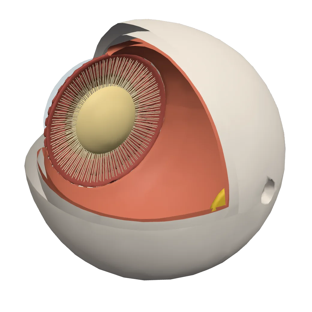
A human eye drawn in 3D by a medical illustrator, cut open at one corner. Tap Show in 3D to turn it and tap its parts.
CorneaThe clear, curved window at the front, where light comes in. It bends the light the most: about two-thirds of the eye’s focusing.
Iris and pupilThe colored ring of muscle in front of the lens. The hole in its middle is the pupil, where light enters: small in bright light, wide in dim light.
Eye lensA clear, flexible convex lens just behind the iris. It hangs in the middle of the ring of ciliary muscle, held all the way round by the suspensory ligaments.
Ciliary musclesA ring of muscle all round the lens. Relaxed, it lets the ligaments pull the lens thin, for far things; contracted, it slackens them and the lens grows thicker, for near ones.
Suspensory ligamentsThin threads from the ring of ciliary muscle to the lens, all the way round, like a wheel’s spokes. They hold the lens, and pulled tight, they pull it thin.
RetinaThe light-sensitive layer lining the back of the eye, with the rods and cones, where the image forms. Cut open here to show the inside.
Fovea (yellow spot)A small spot straight behind the lens, where the cones are packed most tightly: vision is sharpest here.
Blind spotWhere the optic nerve leaves the eye, a little to the nose’s side of the fovea. There are no rods or cones here. (The optic nerve itself is not in this model.)
Sclera (white of the eye)The tough white outer coat of the eye. Cut open here to show the inside.
Eye: K. Browne and H. Schlehlein, HuBMAP Human Reference Atlas (CC BY 4.0), cut open
i. Fibrous tunic
The fibrous tunic (fibrous coat) is the outer, tough coat. It has two parts.
A. Sclera (sclerotic). The white of the eye, about five-sixths of the outer coat. It is dense white fibrous connective tissue, mostly collagen fibers, tough enough to protect the eyeball and to keep its shape against the pressure inside. The six eye muscles are attached to it.
B. Cornea. The front one-sixth of the outer coat: a clear, transparent window that bulges forward like a watch glass. It is the same collagen as the sclera, but its fibers are laid in perfectly regular sheets, which lets light through. Its curved front does most of the eye’s focusing: about two-thirds of the bending of light happens here, at the step from air into the cornea; the lens adds the rest and fine-tunes it.
The cornea has no blood vessels (it is avascular): it gets oxygen straight from the air (through the tear film) and food from the aqueous humor behind it. Because there are no blood vessels to bring the immune system’s cells to it, a cornea from a donor is rarely rejected: corneal transplants are among the most successful transplants of all, and Nepal has an eye bank, at Tilganga, that collects donated eyes.
In the ring where the cornea meets the sclera (the limbus) runs a fine channel, the canal of Schlemm, through which the aqueous humor drains into the veins.
ii. Vascular tunic
The vascular tunic (vascular coat), also called the uvea, is the middle coat, rich in blood vessels and dark pigment. It has three parts.
Eye: traced from Gray’s Anatomy (1918), Fig. 869, H. V. Carter, public domain
A. Choroid. A thin, dark brown, pigmented layer between the sclera and the retina over the back two-thirds of the eye. Its pigment absorbs light that has passed through the retina, so it isn’t reflected back to blur the image (as black paint inside a camera does). It is loose connective tissue full of blood vessels, which feed the outer layers of the retina.
B. Ciliary body. The thickened ring where the choroid continues forward, round the edge of the lens. It has two parts:
- the ciliary processes, folds that secrete the aqueous humor;
- the ciliary muscle, a ring of smooth muscle with circular and meridional (front-to-back) fibers, which changes the lens’s shape when you focus.
From the ciliary body hang the suspensory ligaments (zonules), hundreds of fine fibers attached all round the lens’s elastic capsule. The ciliary body, the ligaments and the capsule together hold the lens in place.
C. Iris. The colored ring that you see through the cornea: a muscular diaphragm that continues forward from the ciliary body and hangs in the aqueous humor in front of the lens. Its color comes from melanin: a lot of it gives brown eyes, as in most Nepalis; very little gives blue. The hole in its middle is the pupil, through which light enters. The iris has two smooth muscles:
- the sphincter pupillae, a ring of circular muscle round the pupil’s edge, which narrows the pupil in bright light (worked by parasympathetic fibers in the oculomotor nerve);
- the dilator pupillae, radial muscle fibers like the spokes of a wheel, which widen it in dim light or fright (worked by sympathetic fibers).
So the pupil changes between about 2 mm and 8 mm across, letting in about sixteen times more light at its widest. The iris divides the space filled with aqueous humor into a large anterior chamber (between cornea and iris) and a narrow posterior chamber (between iris and lens), which meet through the pupil.
iii. Nervous tunic: the retina
The retina (nervous tunic) is the inner, light-sensitive coat. Its outer surface rests on the choroid; its inner surface touches the vitreous humor. It is really a thin sheet of brain: it grows out from the embryo’s brain, and its cells already begin to process the picture before sending it on.
Three places on it have names:
- Macula lutea (yellow spot, area centralis): a yellowish spot in the middle of the back of the retina, directly behind the lens. In its center is a small pit, the fovea centralis, which has only cones, packed tightly: it is the point of sharpest vision. Whatever you look straight at is focused here.
- Blind spot (optic disc): the point, a little to the nose side of the fovea, where the nerve fibers of the retina gather to leave the eye as the optic nerve, and where the retina’s blood vessels enter. It has no rods or cones, so an image falling on it isn’t seen.
- Ora serrata: the wavy front edge of the light-sensitive retina, where it meets the ciliary body.
The ten layers of the retina. Under a microscope a vertical section of the retina shows ten layers, from the outside (the choroid side) in:
| Layer | What is in it | |
|---|---|---|
| A | Pigment cell layer (pigment epithelium) | one layer of cells full of dark pigment, next to the choroid; they absorb stray light and look after the rods and cones |
| B | Rods and cones layer | the light-sensitive outer parts of the rods and cones |
| C | External limiting membrane | a thin boundary between the rods’ and cones’ outer parts and the rest of the retina |
| D | External (outer) nuclear layer | the cell bodies and nuclei of the rods and cones |
| E | External (outer) plexiform layer | a network of fibers where rods and cones synapse with bipolar and horizontal cells |
| F | Internal (inner) nuclear layer | cell bodies of bipolar neurons, horizontal cells and amacrine cells |
| G | Internal (inner) plexiform layer | a network where bipolar cells synapse with ganglion and amacrine cells |
| H | Ganglion cell layer | cell bodies of ganglion cells |
| I | Optic nerve fiber layer | the axons of the ganglion cells, running to the blind spot to leave as the optic nerve |
| J | Internal limiting membrane | the boundary between the retina and the vitreous humor |
Servier Medical Art (smart.servier.com), CC BY 4.0
Notice the strange order: the light-sensitive rods and cones are in the outermost layer, facing away from the light. Light must pass through all the nerve layers first. The signal then goes back the other way: rod or cone → bipolar neuron → ganglion cell → optic nerve, with the horizontal and amacrine cells linking neighbors sideways. This is why the optic nerve has to pass through the retina, making a blind spot. At the fovea the other layers are pushed aside, so the light falls on the cones directly.
Rods and cones, the two kinds of photoreceptor:
| Rods | Cones | |
|---|---|---|
| Shape of the outer part | long, thin rod | short, tapering cone |
| Number in each eye | about 90 to 120 million | about 5 to 6 million |
| Where | everywhere except the fovea; most in the sides of the retina | packed in the fovea, fewer outside it |
| Pigment | rhodopsin (visual purple) | three kinds of iodopsin (photopsins), for red, green and blue light |
| Sensitive to | dim light: night vision | bright light: day vision |
| Color | no: everything in gray | yes: color vision |
| Detail | coarse: many rods share one ganglion cell | fine: in the fovea, about one cone to one ganglion cell |
Rhodopsin is a protein, opsin, holding a small light-catching molecule, retinal, which the body makes from vitamin A. When light hits it, the retinal changes shape, the rhodopsin breaks apart (“bleaches”), and the rod fires a signal. Rebuilding it takes time and vitamin A. That is why, when you walk from bright sunlight into a dark cinema, you see little for the first few minutes (the rods are rebuilding their rhodopsin; full dark adaptation takes about half an hour), and why a shortage of vitamin A causes night blindness.
The cones come in three kinds, each with its own form of opsin (together called iodopsin or photopsins), most sensitive to different colors of light: long-wavelength (red) cones, most sensitive near 564 nm, medium-wavelength (green) cones, near 534 nm, and short-wavelength (blue) cones, near 420 nm. Their pigments are sometimes named erythrolabe, chlorolabe and cyanolabe. Every color you see is the brain comparing how strongly the three kinds respond: this is trichromatic vision.
Which part of the retina has only cones and gives the sharpest vision?
Pick an answer to see why.
Light entering the eye reaches which of these retinal layers last?
Pick an answer to see why.
III. Contents of the eyeball
i. Lens. A transparent, biconvex, elastic body behind the iris and pupil, in front of the vitreous humor. It is about 9 mm across and 4 mm thick, its back surface more curved than its front. It is made of long, clear cells, the lens fibers, packed in layers like an onion’s; as they mature they lose their nuclei and fill with clear proteins (crystallins). A single layer of cuboidal epithelium under its front surface makes new fibers all through life, and the whole lens is wrapped in its elastic capsule. It has no blood vessels. The lens fine-tunes the focus: by changing its curvature (accommodation, below) it brings the image of near or far objects sharply onto the retina. It also separates the aqueous humor in front from the vitreous humor behind.
ii. Aqueous humor. A thin, clear, watery fluid filling the space between the cornea and the lens (the anterior and posterior chambers). It is secreted by the ciliary processes into the posterior chamber, flows through the pupil into the anterior chamber, and drains out through the canal of Schlemm into the blood, being replaced every hour or two. It brings oxygen and food to the cornea and lens, which have no blood vessels, and its pressure, the intraocular pressure, keeps the front of the eye in shape. If it can’t drain, the pressure rises: glaucoma.
iii. Vitreous humor (vitreous body). A clear, thick, jelly-like substance filling the large space between the lens and the retina, four-fifths of the eye’s volume. It is mostly water held in a fine mesh of collagen. It keeps the eyeball in shape, holds the retina against the choroid, and lets light through to the retina. Unlike the aqueous humor, it is formed before birth and not replaced; with age it shrinks and clumps, and the clumps cast shadows on the retina, seen as floaters.
Which fluid drains through the canal of Schlemm?
Pick an answer to see why.
How the eye sees
Vision is the ability to sense the world through the light that enters the eyes, and to make sense of it. It is the eye’s only function. It happens in four steps.
1. Light is focused on the retina. Light from an object is bent (refracted) at the cornea, passes through the aqueous humor, the pupil and the lens, which bends it a little more, crosses the vitreous humor, and comes to a focus on the retina, forming a small, real, inverted image there.
2. The iris controls how much light enters. The pupil widens and narrows (the iris’s muscles), protecting the retina from glare and letting in more light when it is dim. A narrow pupil also sharpens the image, as a small hole does in a camera.
3. The lens adjusts the focus: accommodation. A lens of fixed shape would focus only one distance sharply. The eye changes the curvature of its lens:
- For far objects the ciliary muscle relaxes. The ring of the ciliary body widens, the suspensory ligaments are pulled tight, and they stretch the lens thin and flat: it bends light less.
- For near objects the ciliary muscle contracts. The ring narrows, the ligaments slacken, and the elastic lens springs thicker and rounder: it bends light more.
The ability of the eye to focus on objects at different distances by changing the curvature of its lens is called accommodation. A normal eye sees clearly from far away (its far point, at infinity) down to its near point, taken as 25 cm for an adult (the least distance of distinct vision); a teenager’s eye can focus nearer still, to about 10 cm, as in the model below.
4. The retina turns light into signals, and the brain sees. In the rods and cones light bleaches their pigment, which changes the electrical charge across their membranes (a potential difference). The signal passes through the bipolar cells to the ganglion cells, which fire nerve impulses (action potentials). These run along the optic nerve, through the optic chiasma, to the visual cortex in the occipital lobe at the back of the cerebrum, where they are analyzed: shape, color, movement and depth are worked out, and the brain turns the upside-down images into the upright world you see.
Which is the correct path of light into the eye?
Pick an answer to see why.
When you look up from your book at a distant mountain, the ciliary muscle:
Pick an answer to see why.
Defects of the eye
1. Refractive errors
When the eye can’t bring light to a sharp focus on the retina, the image is blurred: a refractive error. All three are corrected with lenses (glasses or contact lenses), or with laser surgery that reshapes the cornea.
I. Myopia (short-sightedness, near-sightedness). Near objects are clear, but distant objects are blurred. The eyeball is too long, or the cornea and lens too strongly curved, so light from a far object comes to a focus in front of the retina. It is corrected with a concave (diverging) lens, which spreads the light a little before it enters the eye. Myopia usually starts in school years and has become much commoner worldwide as children spend more time indoors and close to screens and books; time outdoors in daylight seems to protect against it.
II. Hypermetropia (hyperopia, long-sightedness, far-sightedness). Distant objects can be seen clearly, but near objects are blurred. The eyeball is too short, or the lens can’t become round enough, so light from a near object would focus behind the retina. It is corrected with a convex (converging) lens.
III. Astigmatism. Blurred or distorted vision at all distances, because the cornea (or the lens) is curved more in one direction than another, like the back of a spoon rather than a ball. Lines running one way are sharp while those at right angles are blurred. It is corrected with a cylindrical lens (curved in one direction only), often combined with a spherical one.
2. Age-related conditions
I. Presbyopia (old-age long-sightedness). From about the age of 40 to 45 the lens gradually loses its elasticity and can no longer round up for near work, so the near point recedes: reading glasses are held farther and farther away. It is corrected with convex reading glasses. A person who also has myopia needs bifocal lenses: the upper part concave for distance, the lower part convex for reading (or varifocals, which change gradually).
II. Cataract. Clouding of the lens, so vision becomes blurred, hazy and dim, as if through frosted glass; the pupil may look milky. It comes mostly with age, sped up by strong sunlight (ultraviolet), smoking, diabetes and eye injury. It is treated by surgery: the cloudy lens is removed and a clear plastic intraocular lens put in its place. Cataract is the leading cause of blindness in Nepal: in the national survey of 2018–2021, it caused about two-thirds (65%) of the blindness among people aged 50 and over.
III. Macular degeneration. Damage to the macula (the yellow spot), mostly in old age, causing loss of central vision: the middle of the view is blurred or dark, while the sides remain.
IV. Floaters (eye floaters). Small specks, threads or cobwebs drifting across the view, especially against a bright sky: shadows of clumps of collagen or cell debris in the vitreous humor. They are usually harmless, but a sudden shower of them, with flashes, can mean the retina is tearing.
3. Other eye conditions
I. Corneal opacity. Loss of the cornea’s transparency, from scarring after an injury, an infection (an ulcer), or a shortage of vitamin A; it can lead to blindness. It is the second leading cause of blindness in Nepal. A corneal transplant can restore sight.
II. Glaucoma. Damage to the optic nerve, usually from raised intraocular pressure when the aqueous humor can’t drain properly. It steals the sides of the view first, so slowly that many people don’t notice until much is lost, and the loss is permanent. Caught early, it is controlled with eye drops or surgery that lowers the pressure. It is the third leading cause of blindness in Nepal.
III. Diabetic retinopathy. Damage to the small blood vessels of the retina by years of high blood sugar in diabetes: they leak and bleed, and fragile new vessels grow, which can lead to blindness. People with diabetes should have their eyes checked every year.
IV. Retinal detachment. The retina peels away from the choroid behind it, cutting it off from its blood supply; the person sees flashes, a shower of floaters, and a dark curtain spreading across the view. It needs urgent surgery.
V. Dry eye. Too few tears, or tears that evaporate too fast (when the Meibomian glands’ oil is short), so the eye feels gritty, burning and tired; long hours at a screen, when we blink less, make it worse. It is treated with lubricating eye drops (artificial tears) and by blinking and resting the eyes often.
VI. Diplopia (double vision). Seeing two images of one object, usually because the two eyes aren’t pointing the same way: a squint, a weak eye muscle, or damage to the nerve that works it (after a head injury, a stroke or with diabetes). Too much alcohol causes it for a while, by upsetting the control of the eye muscles.
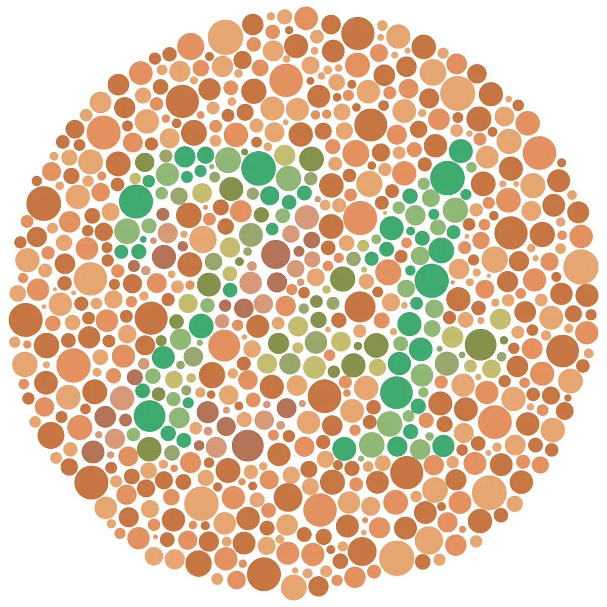
VII. Color blindness. Difficulty telling certain colors apart, most often red from green, because one kind of cone is missing or faulty. Most of it is inherited through a gene on the X chromosome, so it is far commoner in men: among people of European ancestry, about 1 man in 12 but only 1 woman in 200.
VIII. Amblyopia (lazy eye). Reduced vision in one eye because, in early childhood, the brain didn’t learn to use it properly, usually because of a squint or because that eye was much more blurred than the other. Treated early, often by patching the good eye so the weak one must work, it can be corrected; after about age 8 it becomes much harder.
A person can read a newspaper comfortably but cannot read a bus number across the road. The defect, and the lens that corrects it, are:
Pick an answer to see why.
Glaucoma damages vision mainly by:
Pick an answer to see why.
8.6.2 Ear
The ears are stato-acoustic organs: stato- refers to the balance (equilibrium) of the body, acoustic to hearing. Each ear holds two sets of receptors, statoreceptors for balance and phonoreceptors (acoustic receptors) for sound. Like the eyes, the ears are exteroceptors, teloreceptors (they detect distant sources) and organs of special sense; their balance organs also work as proprioceptors.
The two ears are on either side of the head. Each runs inwards from the outside of the head into the temporal bone of the skull, whose hardest part, around the inner ear, is called the auditory capsule. Each is connected to the brain by the vestibulocochlear nerve (the auditory nerve), cranial nerve VIII, which is sensory: its cochlear branch carries hearing and its vestibular branch balance.
Each ear has three parts: the external ear, the middle ear and the internal (inner) ear.
Servier Medical Art (smart.servier.com), CC BY 4.0
I. External ear
The external ear is the part outside, mostly open to the air. It has three parts.
i. Pinna (auricle): the flap we call the ear, one on each side of the head. It is elastic cartilage covered with skin, shaped like a funnel. Its curled outer rim is the helix; the soft hanging part at the bottom, the lobule (ear lobe), has no cartilage, only fibrous tissue and fat with many blood capillaries (which is why it can be pierced, and why a lab takes a drop of blood from it). The pinna’s shape collects sound waves and directs them into the ear canal; its folds also change sounds slightly depending on whether they come from in front, behind or above, which helps you tell where a sound comes from. The muscles that could move it are vestigial in humans: a few people can wiggle their ears slightly, but none can turn them towards a sound as a dog or a horse can.
ii. External auditory meatus (external auditory canal, ear canal): a tube about 2.5 cm long in an adult, gently S-shaped, supported by cartilage in its outer third and by the temporal bone further in. It is lined with skin bearing fine hairs and ceruminous glands, modified sweat glands that make cerumen (earwax). The hairs and wax trap dust, insects and germs, and the wax slowly moves outwards by itself, carrying the dirt with it. The canal carries sound waves to the eardrum.
iii. Tympanum (tympanic membrane, eardrum): a thin, tightly stretched, semi-transparent membrane, almost oval and about 1 cm across, closing the inner end of the canal. Its middle is pulled slightly inwards by the handle of the malleus attached behind it; this point is the umbo. The eardrum separates the external ear from the middle ear.
Functions of the external ear. The pinna collects sound waves and directs them along the canal to the eardrum. The waves of pressure set the eardrum vibrating, at the same frequency as the sound, and it passes the vibrations on to the ossicles of the middle ear. (No nerve impulse is made here: impulses start only in the hair cells of the inner ear.) The hairs and wax of the canal keep out dust and germs.
II. Middle ear
The middle ear lies inside the temporal bone, between the eardrum and the inner ear. It has four parts.
i. Tympanic cavity: a small, irregular chamber, about the size of a pea, filled with air, between the eardrum and the inner ear.
ii. Ear ossicles (auditory ossicles): a flexible chain of three tiny bones, joined by small joints, crossing the tympanic cavity:
- A. Malleus (hammer): the largest, shaped like a hammer; its handle is fixed to the eardrum, its head joined to the incus.
- B. Incus (anvil): the middle one, joined to the malleus on one side and the stapes on the other.
- C. Stapes (stirrup): the smallest bone in the body, about 3 mm long, shaped like a stirrup; its flat footplate fits into the oval window of the inner ear.
iii. Muscles. Two tiny skeletal muscles in the tympanic cavity damp the ossicles when a sound is very loud (the acoustic reflex), protecting the inner ear:
- A. Tensor tympani, attached to the malleus: it pulls the eardrum tighter.
- B. Stapedius, attached to the stapes: the smallest skeletal muscle in the body, about 1 mm long.
A third muscle, the tensor veli palatini (tensor palati) of the soft palate, isn’t in the cavity but at the throat end of the Eustachian tube: it opens the tube each time you swallow or yawn.
iv. Openings. The tympanic cavity has three openings:
- A. Eustachian tube (auditory tube): a tube about 3.5 to 4 cm long joining the middle ear to the nasopharynx (the throat behind the nose); bony near the ear and cartilaginous towards the throat, where it is usually closed. It opens briefly when you swallow or yawn (pulled open by the tensor veli palatini), letting air in or out so that the air pressure in the middle ear stays equal to the pressure outside. Then the eardrum can vibrate freely.
- B. Fenestra ovalis (oval window, fenestra vestibuli): an oval opening in the bone between the middle and inner ear, filled by the footplate of the stapes, held in it by a ring of ligament. Through it the stapes pushes the vibrations into the fluid of the inner ear.
- C. Fenestra rotunda (round window, fenestra cochleae): a smaller, round opening just below the oval window, closed by a thin membrane, the secondary tympanic membrane. When the stapes pushes in at the oval window, this membrane bulges out into the middle ear, letting the fluid of the inner ear (which can’t be squeezed) move.
Functions of the middle ear. The middle ear carries the vibrations of the eardrum to the inner ear through the chain of ossicles and the oval window, without changing their frequency. It also concentrates them. Sound in air barely gets into water: at an open air–water surface about 99.9% of it bounces back. The middle ear gets round this in two ways:
- the eardrum is about 17 times larger in area than the stapes’s footplate, so the force collected over the drum is concentrated onto the small window;
- the ossicles work as a small lever, increasing the force by about 1.3 times.
Together the pressure at the oval window is about 20 times that at the eardrum. The Eustachian tube keeps the air pressure on the two sides of the eardrum equal.
The Eustachian tube connects:
Pick an answer to see why.
Which bone is fixed to the eardrum?
Pick an answer to see why.
III. Internal ear
The internal ear (inner ear, labyrinth) lies deep inside the hardest part of the temporal bone (the periotic or petrous bone). It is called a labyrinth because it is a maze of winding passages. It is really two labyrinths, one inside the other:
i. Bony labyrinth: a set of channels hollowed in the bone, filled with perilymph, a fluid much like the cerebrospinal fluid (rich in sodium).
ii. Membranous labyrinth: a set of membranous sacs and tubes of the same shape, lying inside the bony one and floating in its perilymph. It is filled with a different fluid, endolymph (rich in potassium). The membranous labyrinth is the real stato-acoustic organ: it holds the receptors for both balance and hearing. It has three parts: the vestibule, the semicircular canals and the cochlea.
A. Vestibule
The vestibule is the central part of the membranous labyrinth, two small sacs:
- the utriculus (utricle), the larger, upper sac, into which all three semicircular canals open;
- the sacculus (saccule), the smaller, lower sac, joined to the cochlea.
They are joined by a narrow utriculosaccular duct, from which a long tube, the ductus endolymphaticus, runs back and ends blindly in a swelling, the saccus endolymphaticus, which absorbs endolymph.
Each sac has a patch of sensory cells, a macula (plural maculae). It is made of hair cells, each with a bundle of fine hairs (stereocilia, with one taller kinocilium), and supporting cells. The hairs stick up into a gelatinous otolithic membrane whose top is studded with tiny, heavy crystals of calcium carbonate, the otoliths (otoconia, “ear stones”). When the head tilts, or moves straight forward, back, up or down, the heavy otoliths lag or slide, pulling the gel and bending the hairs. The hair cells then send impulses along the vestibular nerve. The macula of the utriculus lies flat and senses mainly tilting and horizontal movement; that of the sacculus stands upright and senses up-and-down movement.
B. Semicircular canals
The three semicircular canals are hoop-shaped tubes, each about two-thirds of a circle, set at right angles to one another like three faces meeting at the corner of a box, so that every way of turning the head moves fluid in at least one of them:
- the anterior (superior) vertical canal,
- the posterior vertical canal,
- the horizontal (lateral) canal.
Both ends of each canal open into the utriculus. At one end each canal swells into an ampulla, holding a ridge of hair cells, the crista ampullaris (plural cristae). The hair bundles of a crista are buried in a tall cap of jelly, the cupula, which reaches across the ampulla like a swing door. There are no otoliths. When the head turns, the endolymph in the canals lags behind, pushing the cupula and bending the hairs. The cristae sense turning (rotation) of the head, especially its start and stop.
The maculae and the cristae are alike: both are hair cells whose hairs are bent by a gel. They differ in the gel: the macula’s is weighed down with otoliths, so it responds to gravity, tilt and straight-line movement; the crista’s has no otoliths, so it responds to rotation.
C. Cochlea
The cochlea is the organ of hearing: a tube that coils two and three-quarter turns round a central pillar of bone, like the shell of a snail, broad at its base and tapering to its tip. Uncoiled, it would be about 3.5 cm long. Its membranous part, the cochlear duct, is joined to the sacculus by a short ductus reuniens and ends blindly at the tip.
Cut across, the cochlea shows three channels (scalae), side by side along its whole length, separated by two membranes:
a. Scala vestibuli: the upper channel, filled with perilymph. Its floor is Reissner’s membrane (the vestibular membrane). At its base it begins at the oval window, where the stapes pushes on it.
b. Scala tympani: the lower channel, filled with perilymph. Its roof is the basilar membrane. At its base it ends at the round window. The two perilymph channels join at the tip of the cochlea through a small opening, the helicotrema.
c. Scala media (cochlear duct): the middle channel, filled with endolymph, with Reissner’s membrane as its roof and the basilar membrane as its floor. It is the most important of the three, for on its basilar membrane sits the organ of Corti (spiral organ), the receptor organ of hearing, running the whole length of the cochlea.
The organ of Corti is a ridge of cells on the basilar membrane:
- hair cells: one row of inner hair cells (about 3,500), which send almost all the signals the brain hears, and three rows of outer hair cells (about 12,000), which amplify faint vibrations by changing length; from the top of each, a bundle of stiff sensory hairs (stereocilia) projects into the scala media;
- supporting cells: the inner and outer pillar cells (rods of Corti), which lean together to form an arch over a triangular space, the tunnel of Corti; the phalangeal cells (cells of Deiters), which hold the outer hair cells; and the cells of Hensen and cells of Claudius on the outer side;
- the tectorial membrane, a flap of jelly that overhangs the hair cells, the tips of the outer hair cells’ hairs set in it.
From the base of the hair cells, nerve fibers run into the bony core of the cochlea, where they join to form the cochlear nerve.
Servier Medical Art (smart.servier.com), CC BY 4.0
Functions of the internal ear. The vestibule (utriculus and sacculus) and the semicircular canals keep the balance of the body; the cochlea is for hearing.
The organ of Corti is found in the:
Pick an answer to see why.
Which receptor contains otoliths?
Pick an answer to see why.
Functions of the ear
The ear has two functions: equilibrium (balance) and hearing.
I. Equilibrium
Equilibrium, or balance, is the body’s ability to know which way is up and how it is moving, and to keep its position. It is of two kinds:
- i. Static equilibrium: sensing the position of the head relative to gravity when still (is it upright, tilted, lying down?) and straight-line movement. It is the work of the maculae of the utriculus and sacculus.
- ii. Dynamic equilibrium: keeping the body’s position during sudden movements, especially turning of the head. It is the work of the cristae of the semicircular canals.
Mechanism of equilibrium. The utriculus, sacculus and semicircular canals together form the vestibular apparatus, a proprioceptor that keeps track of the head:
- When the head is tilted, or starts or stops moving, the otoliths slide and the endolymph lags, moving the gel of the maculae and the cupula of the cristae.
- The hair cells are bent; this changes their membrane potential and they signal more (or less) to the nerve fibers below them.
- The impulses travel along the vestibular nerve (the vestibular branch of cranial nerve VIII) to the vestibular nuclei in the medulla oblongata and pons, and to the cerebellum.
- These centers compare the signals with what the eyes and the muscles and joints report, and send impulses to the muscles of the neck, body and limbs, and of the eyes, to correct the position and keep the gaze steady.
II. Hearing
Hearing is the sense of sound: speech, music, noises and other vibrations of the air. We hear sounds from about 20 Hz to 20,000 Hz (20 kHz) when young; the top of the range falls with age.
Mechanism of hearing:
- The pinna collects sound waves and the ear canal carries them to the eardrum, which vibrates.
- The vibrations pass across the middle ear through the malleus, incus and stapes, which concentrate them about 20 times in pressure.
- The stapes rocks in the oval window, setting up pressure waves in the perilymph of the scala vestibuli.
- These pressure waves pass through Reissner’s membrane to the endolymph of the scala media and make the basilar membrane vibrate, up and down; the fluid’s movement is released at the round window, which bulges in and out.
- As the basilar membrane moves, the hair cells of the organ of Corti on it move against the tectorial membrane above them, and their hairs are bent. This opens channels in the hair cells, which generate nerve impulses in the cochlear nerve fibers.
- The cochlear nerve (cranial nerve VIII) carries the impulses to the auditory area in the temporal lobe of each cerebral hemisphere, where they are perceived as sound.
How the cochlea tells pitch. The basilar membrane isn’t the same along its length: near the base (by the oval window) it is narrow and stiff, and at the tip (apex) wide and floppy. A sound makes a wave run along it which grows and then dies out; the place where it is largest depends on the frequency: high notes shake the base most, low notes the tip. The brain knows which hair cells are firing, and so which pitch it hears. This was worked out by the Hungarian physicist Georg von Békésy, who won the 1961 Nobel Prize for it. Loudness is told by how many hair cells fire and how fast.
High-pitched sounds make the basilar membrane vibrate most:
Pick an answer to see why.
In which lobe of the cerebrum are sounds perceived?
Pick an answer to see why.
Exercise
A. Tick the correct alternative
- The photoreceptor organ of a vertebrate is the:
Pick an answer to see why.
- An eye is an exteroceptor, a teloreceptor, a special receptor and a:
Pick an answer to see why.
- The human eye is:
Pick an answer to see why.
- The nictitating membrane in humans is:
Pick an answer to see why.
- How many extraocular muscles move each eye?
Pick an answer to see why.
- The size of the pupil is controlled by the:
Pick an answer to see why.
- In the human eye, rod cells contain the pigment rhodopsin. Which pigment is found in cone cells?
Pick an answer to see why.
- The sensory part of the human eye is the:
Pick an answer to see why.
- When a person sees an object, an image forms on the retina. Which sequence is the correct path for light entering the eye?
Pick an answer to see why.
- Vitamin A deficiency causes night blindness. In a night-blind person, which cells fail to work in dim light?
Pick an answer to see why.
- A person who should wear concave (biconcave) glasses doesn’t wear them. Where is the image of a distant object formed in their eye?
Pick an answer to see why.
- A person who can see near objects but not far ones has the defect called:
Pick an answer to see why.
- The Eustachian tube connects the:
Pick an answer to see why.
- Which of these lists the ear ossicles in the correct order, from the eardrum inwards?
Pick an answer to see why.
- The auditory ossicles are in the:
Pick an answer to see why.
- Which of these is concerned with mechanical balance?
Pick an answer to see why.
- The sensory part of the human ear for hearing is the:
Pick an answer to see why.
- The organ of hearing is the:
Pick an answer to see why.
- In which part of the human body is the organ of Corti found?
Pick an answer to see why.
- The organ of Corti is in which part of the internal ear?
Pick an answer to see why.
- The scala tympani is connected with the scala vestibuli by the:
Pick an answer to see why.
- The function of the human ear is:
Pick an answer to see why.
B. Short answer questions
Explain briefly the main structures of the human eye.
Answer
Label the two diagrams, and give the function of each part you label.
Traced from a photo by Exordium, CC BY-SA 3.0, via Wikimedia Commons
Eye: traced from Gray’s Anatomy (1918), Fig. 869, H. V. Carter, public domain
Answer
Write down the defects of the human eye.
Answer
Summarize briefly the external, middle and internal ear of a human.
Answer
Discuss briefly the membranous labyrinth of a human.
Answer
Illustrate and describe briefly the working of the human ear.
Answer
Label the numbered parts of the ear, and write the function of each part.
Servier Medical Art (smart.servier.com), CC BY 4.0
Answer
C. Long answer questions
Describe the structure and function of the human eye, with the help of labeled diagrams.
Answer
Explain in detail, with illustrations, the structure and function of the human ear.
Answer
Printable PDFs
- Study notes 6 pages
- Worksheet 4 pages Answer key
Quiz yourself: Life Processes and the Human Body, Grades 5–12 →
Practice every question in the book, and keep track of the ones you miss →
A companion to your textbook, not a replacement for it: study from the book your school uses, and where the two differ, follow your teacher. Found a mistake? Please tell us.