
In one rainy night, a female bullfrog in a paddy field near Chitwan can lay several thousand eggs, and the male clasped on her back sheds millions of sperms over them. Every one of those cells was made months earlier, inside the two frogs, by the same careful process: a cell with two sets of chromosomes divides so that each egg or sperm gets just one set. Get it wrong, and the embryo dies. Get it right, and a single black-and-white dot in the water becomes a tadpole within a week.
This unit opens the part of the book called developmental biology: how a new animal is built, starting from a single cell. It begins before fertilization, with the making of the two cells that fuse: the sperm and the ovum (egg). The next unit follows the fertilized egg of the frog into a tadpole.
7.1.1 Gametogenesis
In sexual reproduction, a new animal begins as one cell, the zygote, made by the fusion of two gametes (sex cells): a sperm from the father and an ovum (egg) from the mother. The zygote then divides again and again, and its cells grow into the whole many-celled body.
Gametes are made in the gonads, the primary reproductive organs: the testes of a male and the ovaries of a female. The process that makes gametes in the gonads is gametogenesis. It has two forms:
- spermatogenesis, the making of sperms in the testes;
- oogenesis, the making of ova in the ovaries.
The key step in both is meiosis. A body cell of a frog has 26 chromosomes, 13 pairs: it is diploid (2n = 26). Meiosis shares out the chromosomes so that each gamete gets one of each pair, 13 in all: gametes are haploid (n = 13). At fertilization, two haploid gametes fuse, and the zygote has 2n = 26 again. Without that halving, the number would double every generation. So meiosis keeps the number of chromosomes of a species the same, generation after generation.
A frog’s body cells have 26 chromosomes. How many chromosomes does each of its sperms have?
Pick an answer to see why.
7.1.2 Spermatogenesis
Spermatogenesis is the formation of haploid, microscopic, motile sperms in the testes. Each testis is packed with fine coiled tubes, the seminiferous tubules. Their walls are lined by the germinal epithelium, whose cells make the sperms, with large Sertoli cells (nurse cells) among them that hold and feed the developing sperms. Between the tubules, the interstitial (Leydig) cells make the male hormone testosterone.
Spermatogenesis is controlled by hormones: the pituitary gland’s follicle stimulating hormone (FSH) and luteinizing hormone (LH), which makes the Leydig cells release testosterone. In a frog, it speeds up as the breeding season (the rains) comes near.
Spermatogenesis goes through three phases, and then a fourth step that changes the shape of the cells (Figure 15.2).
i. Multiplication phase
The germinal epithelium lining each tubule has diploid cells, the primordial germ cells. They divide again and again by mitosis into a large number of spermatogonia (sperm mother cells), each diploid (2n).
ii. Growth phase
A spermatogonium grows a little, taking in food from the Sertoli cells, and becomes a primary spermatocyte (2n). In this phase its DNA is replicated and proteins are made, ready for meiosis.
iii. Maturation phase
The primary spermatocyte divides by meiosis, which has two divisions:
- Meiosis I (the first maturation division) halves the number of chromosomes: one primary spermatocyte gives two secondary spermatocytes, each haploid (n).
- Meiosis II (the second maturation division) separates the two chromatids of each chromosome: each secondary spermatocyte gives two spermatids (n).
So one primary spermatocyte gives four haploid spermatids, all the same size.
Spermiogenesis
A spermatid is a small, round cell. It is haploid but cannot swim: it has no tail and none of the sperm’s special parts. It is changed into a motile sperm by spermiogenesis (also called spermateleosis). No division happens; the cell is rebuilt:
- the Golgi body forms the acrosome, a cap over the front of the nucleus;
- the nucleus condenses (its DNA packed tightly) and becomes narrow and long;
- the centrioles move behind the nucleus, and the distal one grows the tail’s axial filament: the tail develops, for swimming;
- the mitochondria gather round the start of the tail and form the middle piece;
- most of the cytoplasm is thrown away, as a residual body that the Sertoli cells eat, so the sperm is light and streamlined.
In a frog, the developing sperms lie in bundles, their heads held by a Sertoli cell. The finished sperms are released from the Sertoli cells into the lumen (the central space) of the seminiferous tubule: this release is spermiation. From there they pass through the vasa efferentia and the kidney to the urinogenital duct.
Which of these happens during spermiogenesis?
Pick an answer to see why.
Structure of a sperm
A sperm (spermatozoon) is a small, motile, elongated cell with three parts: a head, a middle piece and a tail (Figure 15.3).
Sperm: Mariana Ruiz Villarreal (LadyofHats), public domain, via Wikimedia Commons
1. Head. The head is the sperm’s broadest part. It is almost all nucleus, haploid, its DNA packed very tightly. At its front is a cap, the acrosome, made from the Golgi body, full of enzymes (sperm lysins: protein-digesting enzymes; in mammals also hyaluronidase, which loosens the cells round the egg). At fertilization, these enzymes digest a path through the egg’s coverings so that the sperm can reach the egg’s membrane.
2. Middle piece. Just behind the head, in the neck, are two centrioles:
- the proximal centriole, next to the nucleus, which enters the egg at fertilization and organizes the spindle of the zygote’s first division (cleavage);
- the distal centriole, which forms the axial filament of the tail.
Around the start of the axial filament, the mitochondria are packed in a spiral. They release the energy (as ATP) that drives the tail, so the middle piece is called the sperm’s power house or energy chamber.
3. Tail. The tail (flagellum) is a long, whip-like part behind the middle piece, many times longer than the head. Its beating drives the sperm through water or fluid. Inside it, the axial filament has nine pairs of microtubules round a central pair: the 9 + 2 pattern of every flagellum and cilium. Most of the tail is its main piece; its thin tip is the end piece.
Sperm: S. J. Holmes, The Biology of the Frog (1906), public domain
A frog’s sperm is built the same way, but its head is long and narrow and its middle piece very short. Frog sperms swim in the pond water itself, round the eggs the female has just laid.
Which part of a sperm gives it the energy to swim?
Pick an answer to see why.
7.1.3 Oogenesis
Oogenesis is the formation of mature, functional female gametes, ova, in the ovaries. It is controlled by hormones too: the pituitary’s FSH makes the follicles grow, and LH brings on ovulation. Like spermatogenesis, it has three phases (Figure 15.5).
i. Multiplication phase
The ovary is lined inside by a layer of cuboidal cells, the germinal epithelium. Its primordial germ cells divide repeatedly by mitosis into a large number of diploid oogonia (egg mother cells).
ii. Growth phase
An oogonium enters meiosis and becomes a primary oocyte (2n). Meiosis then pauses early, in prophase I, and the oocyte grows, for months, stored food pouring into it. This growth is far greater than a spermatocyte’s. The food is yolk, made of proteins and fats: the frog’s liver makes the yolk protein (vitellogenin), the blood carries it to the ovary, and the oocyte takes it in. A layer of follicle cells wraps each growing oocyte, feeding it and making hormones. A frog’s primary oocyte grows from a few hundredths of a millimeter to about 1.5–2 mm across, more than ten thousand times its starting volume.
iii. Maturation phase
When the growth is complete, meiosis continues, with very unequal divisions:
- Meiosis I divides the primary oocyte into two haploid cells of very different size: a large secondary oocyte, which keeps nearly all the cytoplasm and yolk, and a tiny first polar body (polocyte), little more than a nucleus.
- Meiosis II divides the secondary oocyte into a large ovum and a tiny second polar body. The first polar body may also divide into two.
So one primary oocyte gives one functional ovum and two or three polar bodies. The polar bodies take no part in reproduction and soon degenerate. Their job is done: they carried away the extra sets of chromosomes, while the egg kept the food.
In a frog, the egg doesn’t finish meiosis before it is laid. Meiosis I is completed in the ovary, just before ovulation, and meiosis II starts but stops at metaphase II. So a frog lays its eggs as secondary oocytes. Meiosis II is completed only when a sperm enters the egg, and the second polar body is pushed out then (Figure 15.6). Only after that do the egg’s 13 chromosomes form the female pronucleus, which meets the sperm’s.
How many functional ova are made from one primary oocyte?
Pick an answer to see why.
Spermatogenesis and oogenesis compared
| Spermatogenesis | Oogenesis | |
|---|---|---|
| Where | testes (seminiferous tubules) | ovaries |
| Growth phase | short; the cell grows little | long (months); the oocyte grows enormously, storing yolk |
| Meiotic divisions | equal: the cells are the same size | unequal: one large cell and a tiny polar body each time |
| From one primary cell | four sperms | one ovum and two or three polar bodies |
| Extra step | spermiogenesis turns spermatids into sperms | none; in a frog, meiosis II ends only after a sperm enters |
| The gamete | small, motile, with little cytoplasm and no yolk | large, non-motile, with much cytoplasm and yolk |
| Numbers | millions of sperms | a few thousand eggs a season |
Structure of the ovum
A frog lays its eggs as secondary oocytes. Each unfertilized egg (Figure 15.7, A) is a round cell about 1.5–2 mm across. It has no hard shell: it is non-cleidoic (an egg with a shell, like a hen’s, is cleidoic). Its coverings, from the inside out:
- its own plasma membrane;
- just outside that, a thin, clear vitelline membrane (vitelline envelope), made in the ovary; with the plasma membrane under it, it looks like a double layer;
- outside that, a thick coat of jelly (often called albumen), added as the egg passes down the oviduct. It swells in water. It protects the egg from drying out and from germs and fungi, cushions it from knocks, holds the eggs together in a mass, keeps the eggs a little warmer than the water round them, and, tasting bad, makes them less likely to be eaten.
The egg has two halves, with an axis through them:
- The upper half, the animal hemisphere, with the animal pole at its top, is black. Its outer layer is packed with grains of the pigment melanin. It holds the egg’s chromosomes, just under the animal pole, and most of the cytoplasm’s organelles, with little yolk. The dark pigment absorbs sunlight, which warms the egg a little, and shields the chromosomes beneath it from the sun’s ultraviolet light.
- The lower half, the vegetal hemisphere, with the vegetal pole at its bottom, is creamy white and heavy with yolk.
A frog’s egg has a moderate amount of yolk (it is mesolecithal), and the yolk is gathered towards the vegetal pole (the egg is telolecithal). (A hen’s egg is also telolecithal, but with far more yolk; a human egg has almost none.) Eggs are laid in water, stuck together by their jelly, as a mass of spawn.
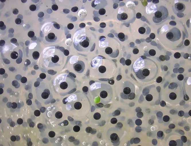
Before fertilization, the egg can lie any way round in its jelly. After a sperm has entered, the egg releases fluid under its vitelline membrane, which lifts off, and the egg can turn freely inside. The heavy yolk then sinks, and every egg turns with its dark animal hemisphere upward, towards the sky and the sun, and its yolky vegetal hemisphere down. Look down on a mass of fertilized frog spawn and you see black dots; look up from below and you see pale ones. (This also hides the eggs from predators: dark from above against the pond’s bottom, pale from below against the bright sky.)
The gray crescent
In the fertilized egg, a band of gray appears on the side opposite the point where the sperm entered (Figure 15.7, B), between the black animal hemisphere and the white vegetal one. This is the gray crescent.
It forms because, after the sperm enters, the egg’s outer layer (the cortex, which carries the pigment) turns by about 30° over the cytoplasm inside it, towards the sperm’s entry point. On the opposite side, the edge of the pigmented cortex moves up, away from the equator, and uncovers a band of the lightly pigmented cytoplasm beneath: the gray crescent.
The gray crescent marks where the dorsal lip of the blastopore will form during gastrulation, and so the future back (dorsal side) of the embryo. Before it appears, the egg has only one axis, animal to vegetal, and is radially symmetrical; the gray crescent gives it a front and back, so the fertilized egg becomes bilaterally symmetrical. More on this in the next unit.
Where does the gray crescent appear in a fertilized frog’s egg?
Pick an answer to see why.
Exercise
The exercise after the next chapter covers this chapter and the next. Its questions on gametogenesis (A1–A7, B1 and B2) are here; the rest are in Unit 16.
A. Choose the best alternative
- What is the final product of spermatogenesis?
Pick an answer to see why.
- In which body organ does gametogenesis occur?
Pick an answer to see why.
- What is the main function of the acrosome in the sperm of a frog?
Pick an answer to see why.
- Which part of the egg in a frog contains the yolk?
Pick an answer to see why.
- Which of the following hormones stimulates ovulation in a frog?
Pick an answer to see why.
- Which of the following structures surrounds the developing oocytes of a frog?
Pick an answer to see why.
- What triggers the completion of oogenesis in a frog?
Pick an answer to see why.
B. Answer the following questions
What is spermatogenesis? Describe it with a chart.
Answer
How does oogenesis occur in a frog? Describe briefly.
Answer
Printable PDFs
- Study notes 3 pages
- Worksheet 4 pages Answer key
Quiz yourself: Life Processes and the Human Body, Grades 5–12 →
Practice every question in the book, and keep track of the ones you miss →
A companion to your textbook, not a replacement for it: study from the book your school uses, and where the two differ, follow your teacher. Found a mistake? Please tell us.