
Leave a slice of bread in a damp tin for a week and a gray fur grows over it, dotted with black pins. A month of monsoon turns a leather shoe green. The same kingdom of living things raises your bread, ferments your raksi, gave the world penicillin, and fills the high pastures of Dolpa with yarsagumba hunters every May. They are the fungi, and most of what they do happens out of sight, as threads too fine to see.
This unit has two chapters. The first is about the fungi: how their bodies are built, how they are classified, and three of them studied closely, the bread mold Mucor, yeast and the mushroom, then the good and harm they do. The second is about the lichens, a fungus and an alga living together so closely that they make one new kind of body.
2.2.1 General introduction
Fungi (singular fungus, Latin for mushroom) are eukaryotic organisms without chlorophyll that feed by absorbing their food. They include the yeasts, the molds and the mushrooms. The branch of biology that studies fungi is mycology (Greek mykes, fungus), and a scientist who studies them is a mycologist. Older books describe fungi as non-green, nucleated, non-vascular thallophytes: plants without roots, stems or leaves. They are no longer counted as plants, but the description still fits them.
Fungi grow wherever there is organic matter to feed on: in soil, air and water, on and in animals and plants, on food, wood, leather and paper. Most prefer warm and damp places, which is why bread goes moldy in the monsoon and stays fresh in a dry winter.
The body: hyphae and mycelium
Except for the yeasts, which are single cells, the body of a fungus is a mass of long, slender, branching threads called hyphae (singular hypha). Each hypha is a fine tube of wall with cytoplasm inside, growing at its tip. The whole network of hyphae is the mycelium. A hypha is too thin to see by itself (usually about 2–10 µm across, a tenth of a hair’s width or less), but the mycelium is the woolly growth on old bread.
Hyphae are of two kinds (Figure 5.1):
- Coenocytic (aseptate) hyphae have no cross walls: one continuous tube of cytoplasm with many nuclei scattered through it, as in Mucor.
- Septate hyphae are divided by cross walls, septa (singular septum), into compartments with one, two or a few nuclei each, as in the ascomycetes and mushrooms. A septum has a pore in the middle, so cytoplasm, and even nuclei, can still flow from one compartment to the next.
The cell wall of fungi is made mainly of chitin, the same tough material as an insect’s outer skeleton, with other sugars (glucans). Their food is stored as glycogen and oil, not as starch.
Nutrition and reproduction
Fungi are heterotrophic: they can’t make their own food. A hypha pours enzymes out into its food and absorbs the dissolved products through its wall. By how they get their food, fungi are:
- saprophytes, which feed on dead organic matter: most molds and mushrooms;
- parasites, which feed on living hosts and harm them: Fusarium, which makes plants wilt; the rusts and smuts of crops;
- symbionts, which live in a partnership that helps both partners: with algae in lichens (2.3), and with the roots of plants as mycorrhiza.
Fungi reproduce in three ways: vegetative (a piece of mycelium breaking off, fragmentation; budding in yeast), asexual (spores formed without any fusion), and sexual (the fusion of two nuclei, then meiosis).
A hypha has no cross walls and many nuclei in one continuous cytoplasm. What is it called?
Pick an answer to see why.
2.2.2 Classification
By the form of the mycelium, the way spores are formed and the fruiting bodies, the older classification divides Kingdom Mycota into four classes. Their names come from the sexual spores or the fruiting body that marks each.
1. Phycomycetes (the algal fungi)
They were called algal fungi because their mycelium resembles some filamentous algae. Their features:
- a.They live in water, on decaying wood in damp places, or as obligate parasites on plants.
- b.The thallus is a mycelium of aseptate, coenocytic hyphae.
- c.Asexual reproduction is by spores formed inside a sporangium: non-motile aplanospores in land species, motile zoospores in water species.
- d.Zoospores have one or two flagella (uniflagellate or biflagellate).
- e.Sexual reproduction is isogamous (similar gametes) or oogamous (a large egg and a small male gamete).
The class is divided into two subclasses:
| Oomycetes | Zygomycetes |
|---|---|
| generally parasites | generally saprophytes, rarely parasites |
| asexual reproduction by motile, biflagellate zoospores | asexual reproduction by non-motile spores; oidia and chlamydospores may form |
| sexual reproduction by gametangial contact: a male gametangium (antheridium) touches a female one (oogonium) and passes its nucleus in | sexual reproduction by gametangial copulation (conjugation): two whole gametangia fuse |
| the fertilized egg is a thick-walled oospore | the fusion gives a thick-walled, resting zygospore |
| examples: Albugo, Plasmopara, Phytophthora | examples: Rhizopus, Mucor |
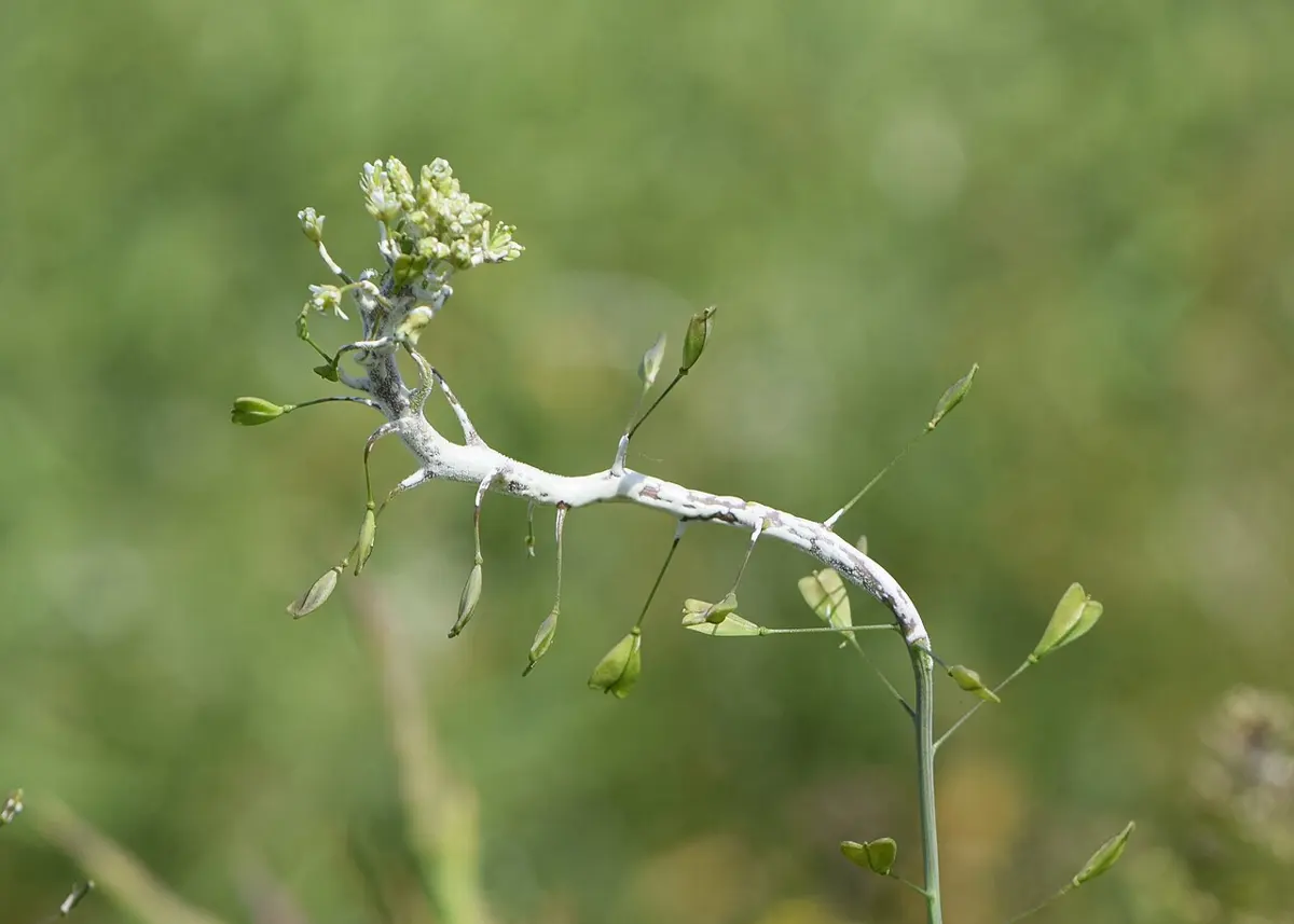
2. Ascomycetes (the sac fungi)
- a.They are called sac fungi because their sexual spores form inside a sac-like cell, the ascus (plural asci).
- b.Most live on land, as saprophytes or parasites.
- c.Most are multicellular, with a mycelium of septate hyphae; the septa have a central pore. Yeast is unicellular.
- d.There is no motile stage anywhere in the life cycle.
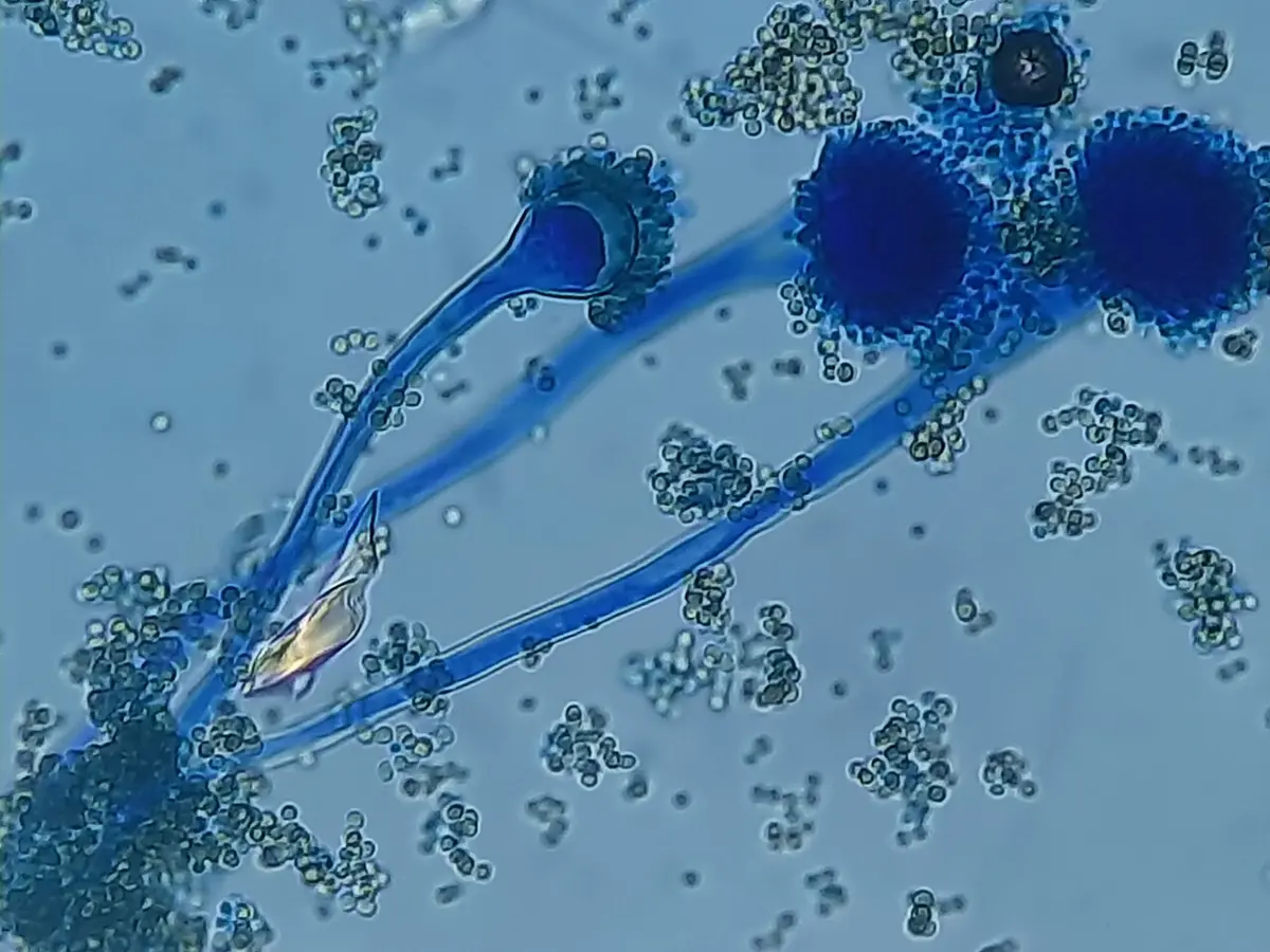
- e.Asexual reproduction is by conidia, spores formed in chains at the tips of special hyphae, the conidiophores. A conidium germinates into a new mycelium.
- f.Sexual reproduction gives ascospores, usually eight (sometimes four), inside each ascus.
- g.Asci may lie free (as in yeast) or grouped in a fruiting body of hyphae, the ascocarp.
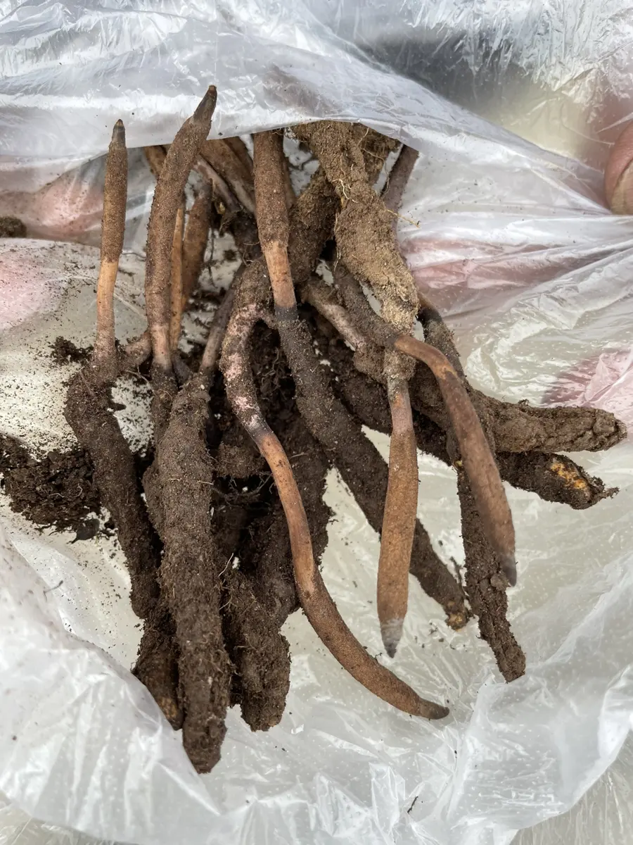
Examples: Saccharomyces (yeast), Penicillium, Aspergillus, Claviceps (ergot of rye), Neurospora (pink bread mold), and the morels (guchhi chyau).
3. Basidiomycetes (the club fungi)
- a.They grow in soil, on logs and tree stumps, and as parasites in living plants.
- b.The mycelium is of branched, septate hyphae. Each septum has a barrel-shaped pore, the dolipore.
- c.The mycelium is of two kinds, primary and secondary.
- d.The primary mycelium is monokaryotic (one nucleus per cell): it grows from a germinating basidiospore, and is short-lived.
- e.The secondary mycelium is dikaryotic (two nuclei per cell, one from each parent): it forms when two primary mycelia of opposite mating types fuse, and it is long-lived; it makes the fruiting bodies.
- f.Vegetative reproduction is by fragmentation; asexual spores are generally absent (except in the rusts and smuts).
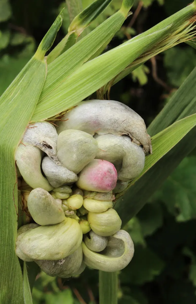
- g.In sexual reproduction, the two nuclei of a dikaryotic cell at the tip of a hypha fuse; the cell becomes a club-shaped basidium, which forms four basidiospores on its outside.
Examples: Agaricus (mushroom), Ustilago (smut), Puccinia (rust of wheat), Polyporus (bracket fungus).
4. Deuteromycetes (Fungi Imperfecti)
- a.They are called imperfect fungi because only their asexual (and vegetative) stage is known. The sexual stage of a fungus is called its perfect stage.
- b.They are saprophytes or parasites.
- c.The mycelium is of well developed, branched, septate hyphae.
- d.Asexual reproduction is by conidia.
- e.Sexual reproduction is absent, or not yet found.
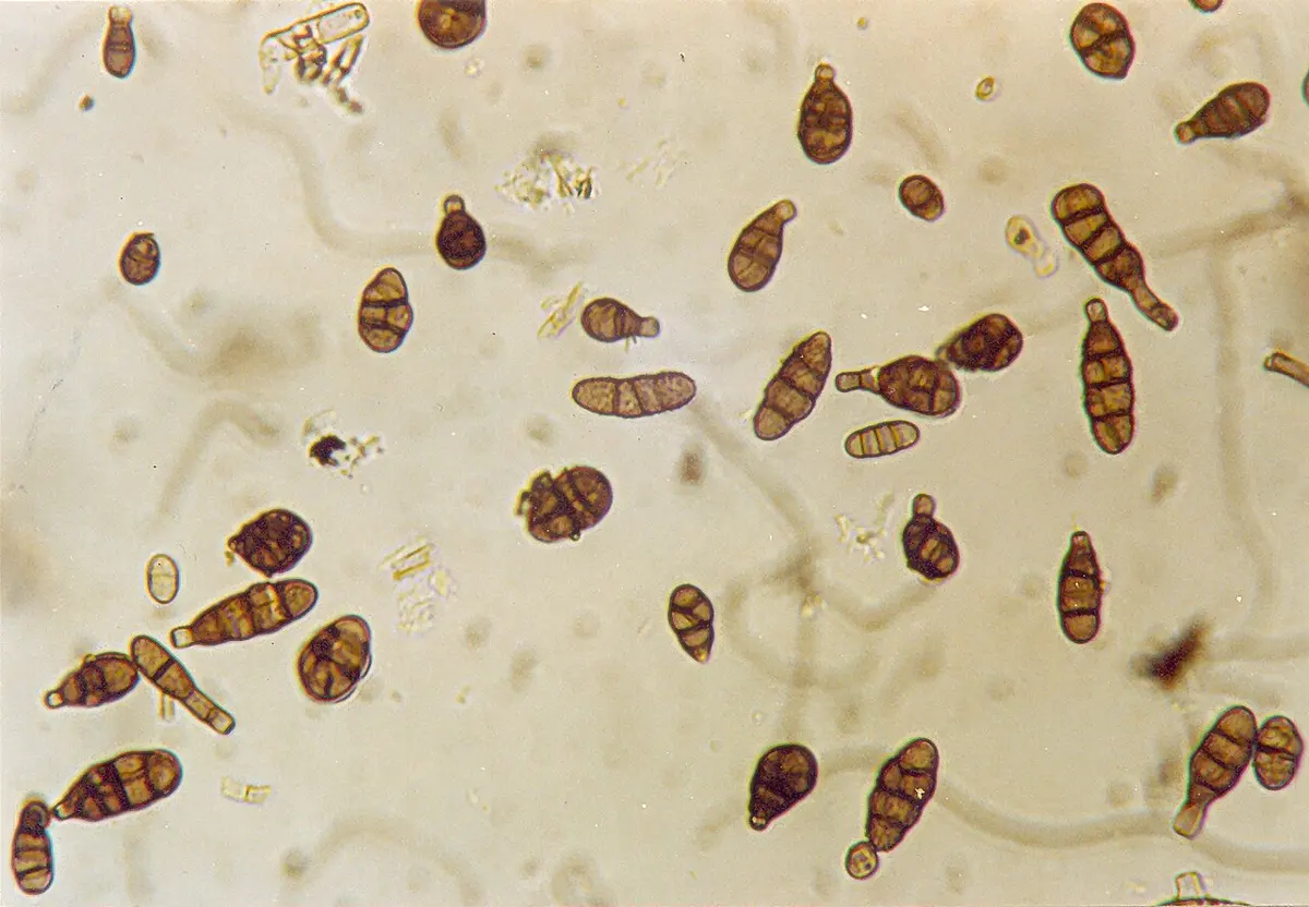
Examples: Alternaria (leaf spot and early blight), Trichoderma, Cladosporium, Fusarium.
In which class do the sexual spores form inside a sac-like cell?
Pick an answer to see why.
A mushroom’s long-lived mycelium has two nuclei, one from each parent, in every cell. What is it called?
Pick an answer to see why.
2.2.3 Mucor
Systematic position
| Rank | Name |
|---|---|
| Kingdom | Mycota (Fungi) |
| Division | Zygomycota |
| Class | Zygomycetes (Phycomycetes in the older system) |
| Order | Mucorales |
| Family | Mucoraceae |
| Genus | Mucor |
Occurrence
Mucor is called the pin mold or bread mold: its sporangia stand on fine stalks like the heads of pins. It is a saprophyte, growing on bread, jam, jelly, cheese, pickles, leather, cattle dung, stored food and soil rich in humus.
Morphology
The vegetative body of Mucor is a mycelium of long, cylindrical, branched, coenocytic hyphae. The mycelium has three kinds of hyphae:
- a.Submerged (subterranean) hyphae: highly branched, smaller, they grow into the substratum and spread inside it. They absorb food and fix the mold in place.
- b.Prostrate hyphae: highly branched, they spread in all directions over the surface of the substratum and help to fix the mold to it.
- c.Aerial hyphae: they grow vertically up from the prostrate hyphae and bear sporangia at their tips, so they are called sporangiophores. They serve in asexual reproduction.
Mucor drawn after Brefeld (Dodel-Port Atlas) and Meyers Konversations-Lexikon (public domain)
Reproduction
Mucor reproduces in three ways: vegetative, asexual and sexual.
1. Vegetative reproduction
By fragmentation: in unfavorable conditions the hyphae break into pieces, mechanically or by decay of parts. When conditions improve, each piece grows into a new mycelium.
2. Asexual reproduction
By three kinds of non-motile structures: sporangiospores, chlamydospores and oidia.
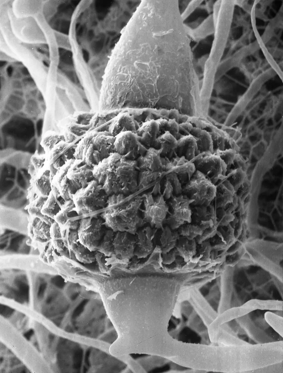
a. Sporangiospores. This is the usual way, in favorable conditions. The tip of a sporangiophore swells into a round sporangium. Inside it, the cytoplasm separates into two regions: an outer, dense, fertile region, the sporangium proper, and an inner, dome-shaped, sterile region, the columella, which is the swollen top of the stalk. The nuclei of the fertile region divide by mitosis many times, and the cytoplasm cuts up into many small, multinucleate sporangiospores, each with its own wall. When they are ripe the columella swells, the thin sporangial wall bursts, and the spores are set free. Carried by air, each spore that lands on a suitable substratum germinates into a new mycelium.
b. Chlamydospores. In unfavorable conditions, the cytoplasm of a hypha gathers at certain places, rounds off, stores a lot of food and grows a thick wall. These resting spores are chlamydospores (Greek chlamys, cloak). In favorable conditions each germinates into a new mycelium.
c. Oidia. When some species of Mucor grow in a liquid rich in sugar, their hyphae become septate, and the short cells round off and separate. These are oidia (singular oidium). They take part in fermentation and multiply by budding, like yeast. Put back on a solid medium, each oidium grows into a mycelium again.
3. Sexual reproduction
Sexual reproduction is by conjugation (gametangial copulation), in unfavorable conditions. Most species are heterothallic: conjugation happens only between two mycelia of different mating types, called the + strain and the − strain (they look alike, so they are not called male and female).
- Hyphae of the + and − strains grow close together and put out short, club-shaped swellings towards each other, the progametangia.
- Their tips meet. A septum forms near the tip of each, dividing it into a basal suspensor and a terminal gametangium.
- The wall between the two gametangia dissolves, and their multinucleate contents fuse: the zygote. Its many nuclei fuse in + and − pairs, so it now has many diploid nuclei.
- The zygote swells, rounds off and secretes a thick, warty, dark wall: the zygospore, held between the two suspensors.
The zygospore rests, sometimes for months. Its dense cytoplasm has many diploid nuclei, organelles and stored food. When it germinates:
- Of its many diploid nuclei, only one stays functional; it divides by meiosis into four haploid nuclei. Three degenerate; the fourth divides many times, so all the nuclei are of the same strain.
- The protoplast swells, bursts the thick outer wall and grows out as an upright germ tube, the promycelium.
- The tip of the promycelium swells into a round germ sporangium, which forms many non-motile, uninucleate, haploid germ spores. Set free, each germinates into a new mycelium.
In Mucor, what is the columella?
Pick an answer to see why.
When are chlamydospores formed in Mucor?
Pick an answer to see why.
2.2.4 Yeast
Systematic position
| Rank | Name |
|---|---|
| Kingdom | Mycota (Fungi) |
| Division | Ascomycota |
| Class | Saccharomycetes (Hemiascomycetes in older books) |
| Order | Saccharomycetales |
| Family | Saccharomycetaceae |
| Genus | Saccharomyces |
Occurrence
Yeasts are saprophytes found all over the world. They grow wherever there is sugar: on ripe and rotting fruit, in sugarcane juice, in the nectar of flowers, on sweets, decaying organic matter and milk products. A few are parasites of plants and animals: Candida albicans causes thrush and other infections in humans.
Morphology
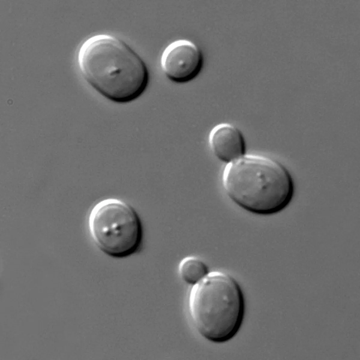
Yeast is a unicellular, non-mycelial fungus, with oval or round cells about 5–10 µm long. When it buds quickly, the cells may stay joined in branched or unbranched chains of up to about a hundred cells; such a temporary chain is a pseudomycelium (false mycelium).
Each cell (Figure 5.11) has:
- a thin cell wall, made mostly of glucans and mannoproteins (sugars and sugar-proteins), with a little chitin;
- the cell membrane inside the wall;
- cytoplasm with mitochondria, endoplasmic reticulum, Golgi bodies and ribosomes, and food stored as glycogen granules and oil droplets;
- one large vacuole, surrounded by its membrane, the tonoplast;
- one nucleus, lying close beside the vacuole, with a nucleolus.
Reproduction
Yeast reproduces vegetatively and sexually.
1. Vegetative reproduction
This happens in favorable conditions, by budding or by fission.
a. Budding. This is the commonest method, in a sugar solution. A small outgrowth, the bud, appears at one end of the parent cell and grows. The nucleus moves towards the bud and divides into two daughter nuclei; one, with part of the cytoplasm, passes into the bud, and the other stays in the parent. The vacuole breaks into small ones while this happens. The bud grows almost to the parent’s size, a wall forms across the narrow neck between them, and the bud separates as a new cell, leaving a bud scar on the parent. When yeast grows fast, the daughter cells bud before they separate, giving a chain of cells, the pseudomycelium.
b. Fission. This is how the fission yeasts (Schizosaccharomyces) multiply. A rod-shaped cell grows longer, its nucleus divides into two, and a cross wall forms in the middle, dividing the cytoplasm into two halves, each with a nucleus. The halves separate and live as two independent cells.
2. Sexual reproduction
Sexual reproduction happens in unfavorable conditions. Yeast forms no sex organs: two vegetative cells of opposite mating types act as gametangia and fuse. This is conjugation. The diploid cell that results, at once or later, becomes an ascus, and meiosis in it forms haploid ascospores. By whether the haploid or the diploid phase of the vegetative (somatic) cells is the main, long-lived one, yeast life cycles are of three kinds.
a. Haplobiontic life cycle (in Schizosaccharomyces octosporus): the haploid phase is dominant.
Two haploid cells of opposite strains come together and act as gametangia. Each puts out an outgrowth towards the other; the outgrowths touch, the wall between them dissolves and forms a conjugation tube. The two nuclei fuse in the tube to form a diploid zygote, which grows and acts directly as an ascus. Its nucleus divides by meiosis into four haploid nuclei, which divide once more by mitosis into eight. Each becomes an ascospore. When the ascus wall bursts, the ascospores are freed and each lives as a haploid, uninucleate vegetative cell. The zygote is the only diploid cell in the cycle.
b. Diplobiontic life cycle (in Saccharomycodes ludwigii, called Saccharomyces ludwigii in older books): the diploid phase is dominant.
In unfavorable conditions a diploid vegetative cell itself becomes an ascus. Its nucleus divides by meiosis into four haploid nuclei of two mating types, which become four ascospores. Still inside the ascus, ascospores of opposite mating types conjugate in pairs. Each zygote puts out a germ tube, which breaks through the ascus wall and grows into a short sprout mycelium, which buds off diploid vegetative cells. The four ascospores are the only haploid cells in the cycle.
c. Haplodiplobiontic life cycle (in Saccharomyces cerevisiae, baker’s and brewer’s yeast): the haploid and diploid phases are equally important.
Two haploid vegetative cells fuse to form a diploid zygote. The zygote behaves as an ordinary vegetative cell and buds into many diploid cells. Each diploid cell can then act as an ascus: its nucleus divides by meiosis into four haploid nuclei, which become four ascospores. Freed from the ascus, the ascospores bud and give haploid vegetative cells, which also live and bud for many generations.
In which life cycle of yeast is the zygote the only diploid cell?
Pick an answer to see why.
2.2.5 Introduction of mushroom
The mushroom is a saprophytic basidiomycete that grows on damp, dead organic matter: humus, animal dung, damp rotting logs, tree trunks and meadows rich in decaying matter. Mushrooms spring up on grasslands and in humus-rich places in the rainy season. The body of a mushroom has two main parts: the mycelium and the basidiocarp.
1. Mycelium (the vegetative body). A mass of profusely branched hyphae grows hidden in the soil or wood, absorbing food. It lives for years. When there is enough food and moisture, its hyphae weave together into a fruiting body.
2. Basidiocarp (the fruiting body). This is the part we call a mushroom. It has a fleshy, cylindrical stalk, the stipe, and an umbrella-like cap on top, the pileus; both are made of tightly interwoven hyphae. When young, the fruiting body is a round button, covered by a thin membrane, the velum (veil). As the pileus grows and opens, the velum tears, and its remains hang round the stipe as a ring, the annulus. On the underside of the pileus are many thin, vertical, radiating plates, the gills (lamellae). The gills are the reproductive part: their surface carries the basidia, which form the spores.
Mushroom: Jan Kops, Flora Batava vol. 10 (1849), public domain
Gill section: G. H. von Schubert, Naturgeschichte des Pflanzenreichs (1887), CC0
Edible and poisonous mushrooms
Some mushrooms are good to eat; some are deadly. Many books give a list of differences like this one:
| Features often said to mark a poisonous mushroom | Features often said to mark an edible one |
|---|---|
| turns green or purple when cut | doesn’t change color when cut |
| bad smell | pleasant smell |
| bitter taste | pleasant taste |
| no insects or worms in it | may have insects or worms in it |
| gills attached to the stalk | gills attached to the cap, free from the stalk |
| annulus usually absent | annulus usually present |
| e.g. Amanita phalloides, Pholiotina rugosa, Cortinarius rubellus | e.g. Agaricus bisporus, Pleurotus ostreatus, Volvariella volvacea |
In the button stage, what covers the young fruiting body of a mushroom, and what does it become?
Pick an answer to see why.
2.2.6 Economic importance of fungi
Fungi do both good and harm in our daily lives.
Useful effects
1. Medicines. Fungi give many drugs. Penicillin, the first antibiotic, comes from Penicillium (P. chrysogenum); other antibiotics such as claviformin also come from Penicillium species. Cyclosporin, from a soil fungus, stops the body rejecting transplanted organs, and statins, which lower blood cholesterol, were first found in fungi. Yeast (Saccharomyces cerevisiae) is a rich source of the B vitamins; the fungus Eremothecium ashbyii is grown to make riboflavin (vitamin B₂), and the mold Blakeslea trispora to make β-carotene, which the body turns into vitamin A. Fungi are also used to make steroid drugs.
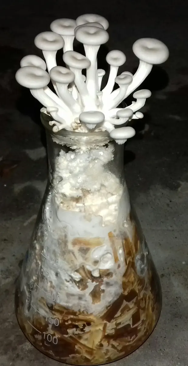
2. Food. People have eaten mushrooms since long ago. Their fruiting bodies have a good amount of protein, vitamins and minerals, and very little fat. In Nepal, the oyster mushroom (Pleurotus, कन्ने च्याउ) and the button mushroom (Agaricus bisporus) are farmed for sale, and wild morels (गुच्छी च्याउ) fetch a high price.
3. Industry. Fungi are used to make many products:
- alcoholic drinks, by fermentation with yeasts such as Saccharomyces; in Nepal the dry starter cake murcha carries the molds Mucor and Rhizopus along with yeasts, which together turn the starch of millet or rice into sugar and then alcohol for jaand, chhyang and raksi;
- bread and other bakery products, with yeast;
- cheese, ripened with Penicillium species (P. roqueforti in blue cheese, P. camemberti);
- enzymes such as amylase, cellulase and invertase;
- organic acids such as citric acid (from Aspergillus niger, for soft drinks), fumaric, lactic and oxalic acids.
4. Soil fertility. Fungi such as Fusarium, Chaetomium and Aspergillus decompose the litter and dead wood of forests. They break down the large molecules of dead bodies (cellulose, hemicellulose, starch, proteins, fats), and the white-rot fungi even lignin, which almost nothing else can digest. The minerals are released back into the soil, so its fertility increases.
5. Plant nutrition. In a mycorrhiza (fungus-root), fungal hyphae live on or in a plant’s roots. The hyphae reach far into the soil and absorb water and minerals, especially phosphate, for the plant, which gives the fungus sugar in return. Most land plants, including the sal and pine of Nepal’s forests, depend on mycorrhizae.
6. Plant hormones. The plant hormone gibberellin was discovered in a fungus. In Japan, rice seedlings infected with Gibberella fujikuroi grew absurdly tall and fell over, the “foolish seedling” (bakanae) disease; in 1926 Eiichi Kurosawa showed that the fungus made a substance that caused it. Gibberellins are still made from fungi for use in farming.
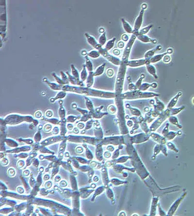
7. Biological control. Some fungi are used to control pests and diseases without chemical poisons. Beauveria bassiana, a soil fungus, infects and kills insects such as beetles, termites and caterpillars. Trichoderma species attack the fungi that rot crop roots, and are sold to Nepali farmers to mix into the soil.
Harmful effects
Fungi harm people directly and indirectly: by diseases of plants, humans and animals, and by spoiling food and goods.
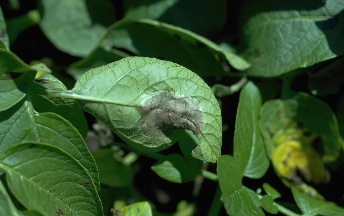
1. Plant diseases. Fungi and fungus-like organisms cause many diseases of crops: late blight of potato and tomato (Phytophthora infestans, a water mold); blast of rice (Magnaporthe oryzae); rusts of wheat (Puccinia); red rot of sugarcane (Colletotrichum falcatum); wart disease of potato (Synchytrium endobioticum); damping off of seedlings (Pythium, a water mold); and club root of cabbage (Plasmodiophora brassicae, a fungus-like protist).
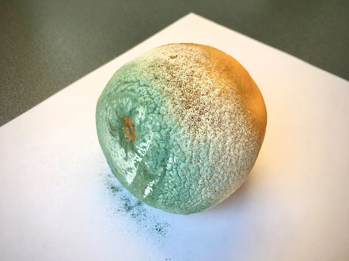
2. Rotting of fruit and food. Poorly stored crops and fruit are attacked by molds, with great losses: Penicillium causes the green and blue molds of oranges and apples, Aspergillus niger black mold, and Aspergillus flavus a yellow-green mold on grains and groundnuts. A. flavus also makes aflatoxins, poisons that damage the liver and can cause cancer. Fungi also spoil vegetables, milk products and meat.
3. Human and animal diseases. Some fungi are parasites of humans and animals: ringworm and athlete’s foot (Trichophyton, Microsporum) infect skin, hair and nails; Candida causes thrush; Aspergillus can infect the lungs. Water molds such as Saprolegnia attack the eggs and gills of fish, a serious problem in fish farms.
4. Damage to clothes. Fungi grow on damp clothes and shoes and rot them. Natural fibers (cotton, wool, silk) are attacked more than synthetic ones.
5. Damage to paper and wood. Many fungi destroy the paper of books and newspapers kept in damp places. Bracket fungi such as Polyporus rot timber and the wood of buildings.
6. Mushroom poisoning. Poisonous mushrooms cause vomiting, diarrhea, stomach pain, and in the worst cases liver failure and death. Nepal’s newspapers report deaths every monsoon.
Exercise: fungi
A. Choose the correct option
- Which group of fungi are called the club fungi?
Pick an answer to see why.
- Which nutrient is commonly obtained from yeast, making it a valuable dietary source?
Pick an answer to see why.
- In fungi, in what form is reserve food mainly stored?
Pick an answer to see why.
- “The mode of nutrition in fungi is mainly saprophytic.” Which interpretation of this statement fits best with the role of fungi in ecology and the cycling of nutrients?
Pick an answer to see why.
- What does the “perfect stage” of a fungus mean?
Pick an answer to see why.
- Which of the following is not a mode of asexual reproduction?
Pick an answer to see why.
- How does Mucor contribute to the ecosystem?
Pick an answer to see why.
Mushroom: Jan Kops, Flora Batava vol. 10 (1849), public domain
- Look at the mushroom in Figure 5.21. Which gives the correct names of the parts A, B, C and D?
Pick an answer to see why.
B. Answer the following questions
Define the following terms: a. mycelium, b. hyphae, c. ascus, d. coenocytic hyphae.
Answer
Compare and contrast the diplobiontic and haplobiontic life cycles of yeast, focusing on their key characteristics, reproductive strategies and ecological implications.
Answer
Justify the statement “Deuteromycetes are known as Fungi Imperfecti”.
Answer
A grocery store has a problem with strawberries spoiling. Suggest measures the store can take to reduce the spoiling.
Answer
Describe the structure of a mushroom with a labeled diagram.
Answer
Describe the vegetative structure of Mucor with a labeled diagram.
Answer
Discuss the ecological importance of fungi and their interactions with other organisms in ecosystems.
Answer
Look at Figure 5.22 and answer the questions.
- a.Name the organism shown in the diagram.
- b.Label A, B and C.
- c.Write the function of B and C.
- d.Mention any one ecological importance of this organism.
Answer
Several pairs of mushroom species closely resemble each other, one poisonous and the other edible. Make a table comparing the features that tell them apart, and discuss the dangers of mistaking one for the other.
Answer
2.3.1 General introduction
A lichen is an organism made of a fungus and an alga so closely associated that they appear to be a single organism. So a lichen is a dual or composite organism. The fungal partner is the mycobiont; the photosynthetic partner is the phycobiont (a green alga in most lichens; in about a tenth of them it is a cyanobacterium, then also called the cyanobiont).
In the lichen’s body, the thallus, the fungus makes up most of the volume. It gives the lichen its shape and much of its color, shelters the alga from drying out and strong light, and absorbs water, minerals and carbon dioxide for both. The alga makes food by photosynthesis, and the food is shared by both partners. Neither can grow like this alone: on its own the fungus would starve, and the alga would dry up on a bare rock.
The study of lichens is lichenology. The Swedish botanist Erik Acharius (1757–1819), who first classified lichens as a group of their own, is called the father of lichenology. Over two thousand years earlier, the Greek Theophrastus, the “father of botany”, was the first to describe lichens, and some books give the title to him; mentioning both, with what each did, is safe in an exam.
Lichen: Encyclopaedia Britannica (1911), public domain
2.3.2 Characteristic features of lichens
- They are dual organisms: a symbiotic association of an alga (or cyanobacterium) and a fungus.
- They usually grow in moist, cool places with moderate temperatures, on the bark of trees, leaves, dead logs, bare rocks and walls. Many survive drought and cold by drying out completely and reviving when wet.
- They grow very slowly, often only a few millimeters a year, and can live for hundreds of years.
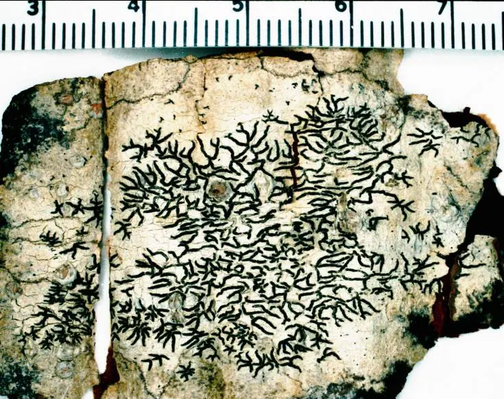
- The body is a thallus: irregular in shape, and often brightly colored, mostly by pigments and acids made by the fungus. By the form of the thallus and how it is attached to the substratum, lichens are of three types:
- crustose: crust-like, closely attached to and partly embedded in the substratum, so that it can’t be lifted off whole (Graphis, Lecanora);
- foliose: flat and leaf-like, with lobes, attached at points and easily lifted (Physcia, Parmelia, Xanthoria);
- fruticose: branched and shrubby, upright or hanging, attached only at its base (Usnea, Cladonia).
- They are very sensitive to air pollution, and are used as indicators of it.
- They reproduce by vegetative, asexual and sexual methods: by fragments of the thallus and tiny powdery packets of fungus and alga together (soredia) that blow away; the fungus also forms its own spores.
Examples: Graphis, Physcia, Usnea.
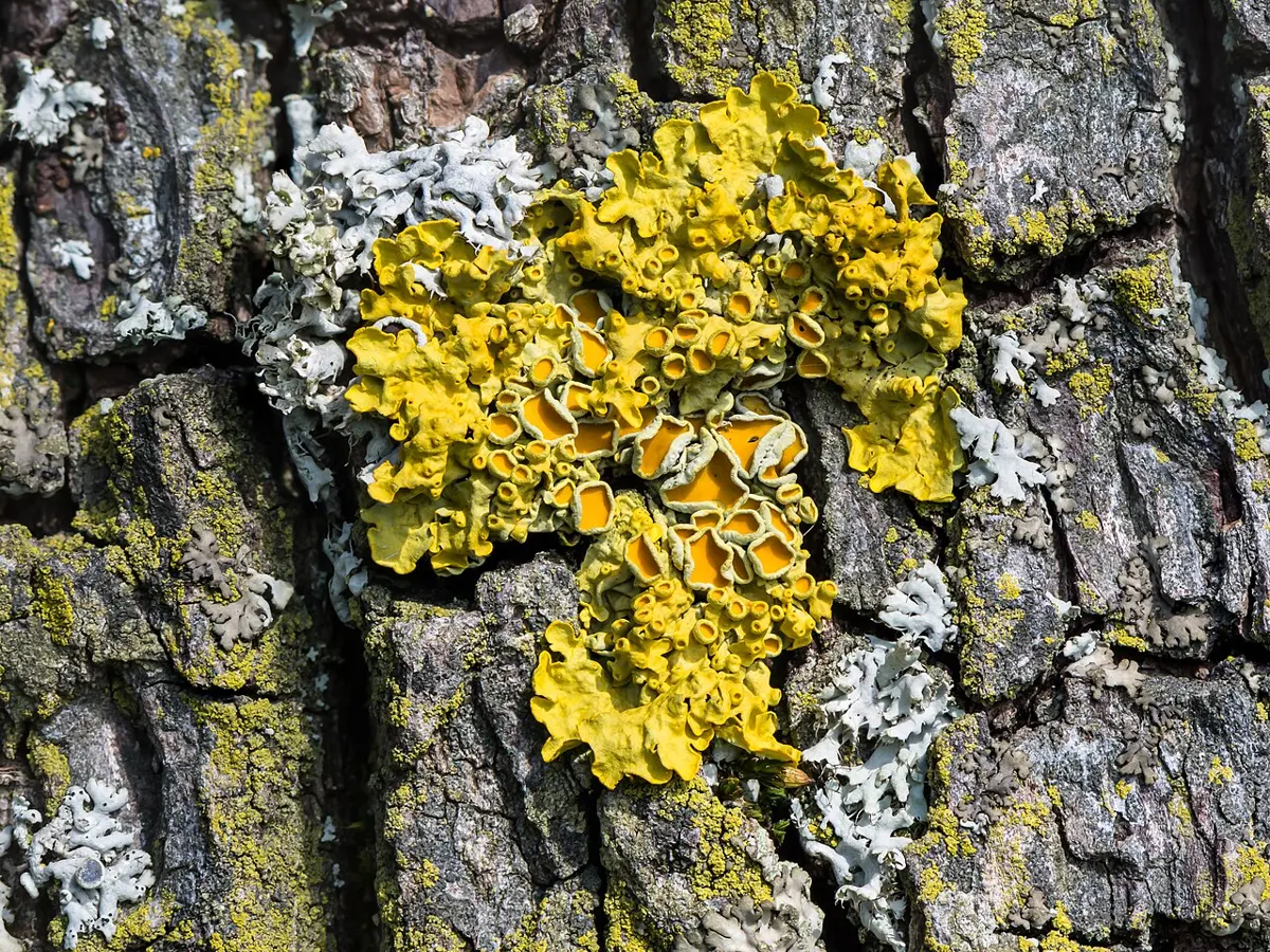
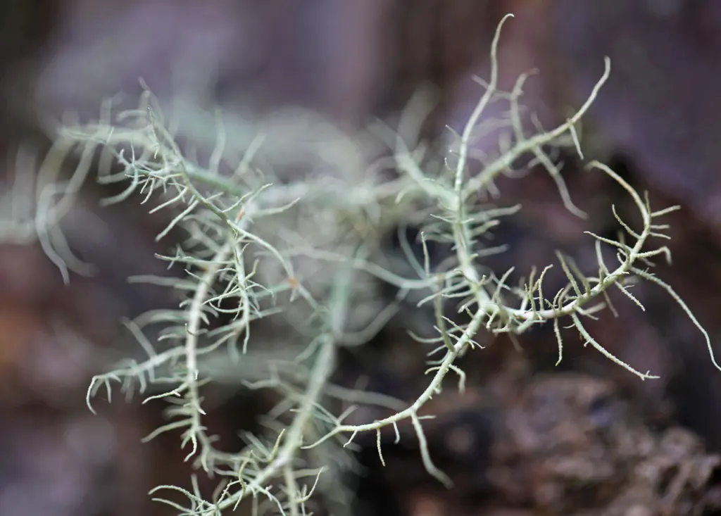
Which kind of lichen is shrubby or hanging, attached to the substratum only at its base?
Pick an answer to see why.
2.3.3 Economic importance of lichens
Lichens have both useful and harmful effects.
Useful effects
1. Food. Lichens feed many small animals: mites, slugs, snails and caterpillars. In the Arctic, reindeer live through the winter on “reindeer moss”, the lichen Cladonia rangiferina. Some lichens are eaten by people: Parmotrema (stone flower) is a spice in garam masala.
2. Medicine. Lichens have long been used in folk medicine: lung lichen, Lobaria pulmonaria, for lung diseases, because it looks like lung tissue, and the yellow Xanthoria parietina for jaundice. These uses were chosen by the looks of the lichen, and there is no good evidence that they work. Lichens do make real antibiotics, though: usnic acid, from Usnea and others, kills many bacteria and is used in some ointments.
3. Pioneers of ecological succession. Lichens are often the first organisms to colonize bare rock, where nothing else can grow. They hold dust and water, and the acids they release slowly crumble the rock. Their dead remains mix with the rock grains into the first thin soil, in which mosses, then grasses, can take root.
4. Bioindicators of air pollution. Lichens are very sensitive to pollutants such as sulphur dioxide. They absorb water and whatever it carries straight through their surface, they have no way of getting rid of the poisons, and they live for many years, so pollution builds up in them. As air pollution increases, first the shrubby kinds disappear, then the leafy ones, and in the worst air only a few crusts, or none, survive. So the lichens present on trees tell how clean the air is.
5. Industry. Lichens give dyes (the orchil purples, and the tweed dyes of Scotland), litmus, the acid-base indicator (from Roccella), perfumes (oakmoss, Evernia prunastri, in many famous perfumes), cosmetics and even alcohol.
Harmful effects
- Lichens growing on the walls of buildings damage the plaster and spoil the look of the walls.
- In the dry season some lichens become so dry that they catch fire easily and help forest fires spread.
- The acids of lichens etch the surface of glass and marble and reduce their value; they damage old stone statues and stained-glass windows.
- Some lichens grow on the evergreen leaves of crops such as coffee, tea and rubber, shade them and cause early leaf fall.
Exercise: lichens
A. Choose the correct option
- Which pair of organisms forms the symbiotic relationship in a lichen?
Pick an answer to see why.
- What is the field of study that focuses on lichens?
Pick an answer to see why.
- Which of the following describes the body of a lichen?
Pick an answer to see why.
- In the symbiosis of a lichen, what does the alga mainly provide?
Pick an answer to see why.
- Why are lichens often called pioneers in the development of ecosystems?
Pick an answer to see why.
- Which of these harmful effects is associated with lichens?
Pick an answer to see why.
B. Answer the following questions
Mention the characters of lichens.
Answer
Lichens are called dual organisms. Give reasons.
Answer
Define the terms “phycobiont” and “mycobiont” and describe their roles in the lichen symbiosis.
Answer
What are the distinguishing characteristics of fungi and lichens?
Answer
Imagine you are a researcher studying air quality in a city with much industry. How could you use lichens as bioindicators to assess air pollution? Discuss the characteristics of lichens that make them sensitive to pollutants, and explain how changes in lichens could show changes in air quality.
Answer
Write short notes on the economic importance of lichens.
Answer
Printable PDFs
- Study notes 4 pages
- Worksheet 5 pages Answer key
Quiz yourself: Classification of Living Beings, Grades 5–12 →
Practice every question in the book, and keep track of the ones you miss →
A companion to your textbook, not a replacement for it: study from the book your school uses, and where the two differ, follow your teacher. Found a mistake? Please tell us.