
Half an hour after you eat a plate of dal bhat, the sugar in your blood has risen by about half. Within two hours it is back where it started, although you did nothing. In the meantime a few thousand tiny clusters of cells in your pancreas noticed the rise, poured a protein into the blood, and every muscle and liver cell that the protein reached opened its doors to sugar. If you are suddenly frightened, a gland on top of each kidney does something similar in a few seconds, and your heart races before you know why. A pea-sized gland under your brain, steered by the brain itself, decides how fast you grow, when you reach puberty, and how much water your kidneys keep.
These are the endocrine glands, and their chemical messengers are the hormones. This unit visits them one by one: where each gland is, how it is built, what its hormones do, and what happens when it makes too little or too much.
Glands
A gland is a cell, or a group of cells, usually of epithelial tissue, that makes and releases a useful substance: an enzyme, a hormone, mucus, sweat, milk. The body’s glands together are its glandular system. By how they release what they make, there are three kinds.
| Kind | How it releases its secretion | Where the secretion acts | Examples |
|---|---|---|---|
| Exocrine (ducted) glands | through a duct, onto a surface or into a cavity | near where it is made | salivary glands, sweat glands, liver, gastric glands, tear glands |
| Endocrine (ductless) glands | straight into the blood (through the tissue fluid) | usually far away | pituitary, thyroid, adrenal glands |
| Heterocrine (mixed) glands | both: one part through a duct, another into the blood | both | pancreas, testes and ovaries |
An exocrine gland’s secretion is not always an enzyme: sweat, tears, mucus, bile and milk are exocrine secretions too. The pancreas is heterocrine because most of it pours digestive juice down the pancreatic duct while its islets put hormones into the blood; a testis sends sperm down a duct but testosterone into the blood.
The study of the endocrine glands and their hormones is endocrinology. Too much of a hormone (hypersecretion) or too little (hyposecretion) causes disorders, and most of this unit is about those.
Body and glands: BodyParts3D (CC BY 4.0)
Which of these is a heterocrine gland?
Pick an answer to see why.
Hormones
The word hormone was introduced by Ernest Starling in 1905, from the Greek hormao, “I set in motion” or “I arouse”. A hormone is a chemical messenger: a substance made by an endocrine gland (or by scattered endocrine cells), released into the blood in response to a change inside or outside the body, and carried to target cells elsewhere, whose activity it changes. A cell is a target only if it has a receptor that fits the hormone, which is why a hormone carried everywhere acts only in a few places.
Chemical nature of hormones
| Kind | What it is made of | Examples |
|---|---|---|
| Steroid hormones | made from cholesterol; fat-soluble | testosterone (testes), estrogen and progesterone (ovaries, placenta), aldosterone, cortisol and corticosterone (adrenal cortex) |
| Amino acid derivatives | made from a single amino acid: tyrosine | thyroxine (T₄) and T₃, adrenaline and noradrenaline |
| Peptide hormones | short chains of amino acids | oxytocin, vasopressin (ADH), MSH, ACTH, parathormone, calcitonin |
| Protein hormones | long chains | insulin, glucagon (pancreas), growth hormone, prolactin (anterior pituitary) |
| Glycoproteins | proteins with sugar chains attached | TSH, FSH, LH (anterior pituitary); erythropoietin (kidney); inhibin (testes and ovaries) |
| Eicosanoids | made from a fatty acid, arachidonic acid | prostaglandins |
Peptides, proteins and glycoproteins are one family: chains of amino acids of different lengths, so many books put them together as proteinaceous hormones.
Properties of hormones
- They act in very small amounts: a few billionths of a gram in a liter of blood is enough.
- Most are small molecules compared with enzymes, so they pass easily through the walls of capillaries.
- They are released into the blood (into the veins leaving the gland) and act far away.
- Peptide and protein hormones dissolve in the plasma. Steroid and thyroid hormones do not dissolve well in water, so most of each is carried bound to proteins of the plasma.
- They are not catalysts: a hormone carries a message. It binds to its receptor, and the target cell changes what it does: switches genes on or off, makes an enzyme work faster or slower, opens a channel.
- Once their work is done they are broken down (mostly in the liver) or excreted in urine, so the gland must keep making them; this is how the body can turn a signal off.
- Many are made first as a larger, inactive prohormone and cut to size: insulin is cut from proinsulin.
- The body’s own hormones do not normally set off its immune system.
Functions of hormones
Hormones control: metabolism (how fast cells use food and energy); homeostasis (keeping blood sugar, salt, water and calcium steady); growth, maturation and repair; the sex characters and reproduction; the work of other endocrine glands; and the body’s adaptations to stress, cold, hunger and danger.
Hormones and enzymes
| Hormones | Enzymes | |
|---|---|---|
| Made by | endocrine glands | exocrine glands, and all cells |
| Where they act | carried by blood to a distant site | usually near where they are made, or inside the cell that made them |
| Size | mostly small molecules | large molecules |
| Chemical nature | steroids, proteins, peptides, amino acid derivatives | nearly all proteins |
| After acting | broken down, so they must keep being made | not used up: one enzyme molecule works again and again |
| Speed | some act in seconds (adrenaline), some over days (growth hormone) | act very fast |
| Effect | may speed a process up or slow it down | speed up a reaction |
Which is true of hormones but not of enzymes?
Pick an answer to see why.
Thyroxine and adrenaline are both made from:
Pick an answer to see why.
Hypothalamus
The hypothalamus is a small region at the base of the brain, the floor of the diencephalon of the forebrain, about the size of an almond, just above the pituitary gland. Like the rest of the nervous system it develops from the ectoderm of the embryo. It is where the nervous system and the endocrine system meet: its nerve cells make hormones, and through them it controls the pituitary gland, which controls most of the others. So it is called the supreme commander of endocrine regulation. (Its place in the brain: Diencephalon.)
Its neurons are grouped into nuclei, clusters of nerve cells each with its own job, in three regions from front to back.
1. Anterior region (supra-optic region)
Above the optic chiasma. Its main nuclei are the supra-optic and paraventricular nuclei, with smaller ones round them. Most of the hypothalamus’s hormones are made here:
- Corticotropin-releasing hormone (CRH): makes the anterior pituitary release ACTH.
- Thyrotropin-releasing hormone (TRH): makes the anterior pituitary release TSH.
- Gonadotropin-releasing hormone (GnRH): makes the anterior pituitary release the reproductive hormones FSH and LH.
- Oxytocin: for childbirth and the release of breast milk; it also affects behavior: bonding between mother and baby, trust, recognition, sexual arousal.
- Vasopressin, or antidiuretic hormone (ADH): controls how much water the kidneys keep.
- Somatostatin: stops the anterior pituitary releasing some of its hormones, such as growth hormone and TSH.
The anterior region also helps to keep body temperature down (by sweating) and keeps the body’s daily circadian rhythm in time with day and night.
2. Middle region (tuberal region)
Its main nuclei are the ventromedial nucleus, which helps control appetite (it signals “full”), and the arcuate nucleus, which makes growth hormone-releasing hormone (GHRH), telling the pituitary to release growth hormone.
3. Posterior region (mammillary region)
Its main nuclei are the posterior hypothalamic nucleus, which keeps the body warm in the cold by causing shivering and stopping sweating, and the mammillary nuclei (in the mammillary bodies), which are part of the circuits of memory.
Which part of the hypothalamus makes oxytocin and ADH?
Pick an answer to see why.
Pituitary gland (hypophysis)
The pituitary gland hangs from the base of the brain just below the hypothalamus, joined to it by a short stalk, the infundibulum. It sits in a hollow of the sphenoid bone in the floor of the skull, the sella turcica (Latin for “Turkish saddle”, from its shape). It is oval, reddish-brown, about the size of a pea: roughly 1.3 cm across and 0.5 g. It is often called the master gland, because its hormones control the thyroid, the adrenal cortex and the gonads; but it is itself controlled by the hypothalamus, and the two work as one unit, the hypothalamo-hypophyseal axis.
Structure
The pituitary has three lobes:
- the anterior lobe (pars distalis), the largest;
- the middle lobe (pars intermedia), a thin strip of tissue between the other two, tiny in adult humans;
- the posterior lobe (pars nervosa).
It has two origins, from two different parts of the ectoderm, and they stay different. In the embryo an upgrowth from the roof of the mouth cavity, Rathke’s pouch, rises to meet a downgrowth from the floor of the brain. Rathke’s pouch becomes the anterior and middle lobes, together called the adenohypophysis (the glandular part, about 75% of the gland); the downgrowth of the brain becomes the posterior lobe, the neurohypophysis (the nervous part, about 25%).
Servier Medical Art (smart.servier.com), CC BY 4.0
- Adenohypophysis. Its cells are named by how they stain: acidophils (the somatotrophs, which make growth hormone, and the lactotrophs, which make prolactin), basophils (the thyrotrophs, corticotrophs and gonadotrophs) and chromophobes, which hardly stain. The releasing hormones of the hypothalamus reach it through the hypophyseal portal veins (a portal vein runs from one net of capillaries to another, without passing through the heart).
- Neurohypophysis. It is made of the axons of neurosecretory cells whose cell bodies are in the hypothalamus, ending in axon terminals among large branched glial cells, the pituicytes. The two hormones made by those cells, oxytocin and ADH, travel down the axons and are stored in swellings, the Herring bodies, and in the terminals until a nerve impulse releases them into the blood. So the posterior lobe makes no hormone of its own: it is a store and a release point.
A. Hormones of the adenohypophysis
Most of them are tropic (trophic) hormones: hormones that make another endocrine gland work. Growth hormone and prolactin are the exceptions: they act on body tissues directly.
- Thyroid-stimulating hormone (TSH), or thyrotropin, from the thyrotrophs, makes the thyroid gland grow and secrete T₃ (triiodothyronine) and T₄ (thyroxine).
- Adrenocorticotropic hormone (ACTH), from the corticotrophs, makes the adrenal cortex secrete its steroid hormones, above all the glucocorticoids, and also the sex corticoids.
- Gonadotropins, from the gonadotrophs:
- Follicle-stimulating hormone (FSH): in a woman, makes the ovarian follicles grow and ripen and secrete estrogen; in a man, acts on the seminiferous tubules to support the making of sperm.
- Luteinizing hormone (LH): in a woman, triggers ovulation and turns the empty follicle into the corpus luteum, which makes progesterone; in a man, makes the Leydig (interstitial) cells of the testis make testosterone (so in men it is also called ICSH, interstitial cell-stimulating hormone).
- Prolactin (PRL), also called lactogenic or luteotropic hormone (LTH), from the lactotrophs: makes the mammary glands grow during pregnancy and make milk after the birth. Too much prolactin causes galactorrhea (milk from the breasts when not breastfeeding), amenorrhea (no periods) and infertility in women, and in men gynecomastia (enlarged breasts) and infertility.
- Melanocyte-stimulating hormone (MSH): in fishes and amphibians it is made by the middle lobe and darkens the skin by spreading the pigment in its pigment cells (melanophores); that is how a frog matches its background. In adult humans the middle lobe is barely present; MSH is made in small amounts by the skin and the brain instead, where it helps the skin tan and curbs appetite.
- Growth hormone (GH), or somatotropin, from the somatotrophs: makes the body grow by making cells divide and specialize. It controls the growth of bones, muscles and other tissues, and it affects the metabolism of carbohydrate, protein and fat: it raises blood sugar and makes the body burn fat and build protein. It is released mostly in deep sleep.
Disorders of growth hormone
Too little in childhood:
- Pituitary dwarfism (ateliosis): too little GH in infancy and childhood stunts growth. The person is short but well-proportioned, with normal intelligence (unlike a cretin). It is treated with injections of growth hormone, now made by genetically engineered bacteria.
- Simmonds’ disease (pituitary cachexia): when the whole anterior pituitary is destroyed in an adult, by a tumor or by heavy bleeding in childbirth, all its hormones fail, growth hormone among them. The person grows thin and weak, ages early, the sex organs waste, and muscles and connective tissue degenerate.
Too much:
- Gigantism: too much GH in childhood, before the growth plates of the long bones close: the person grows very tall, with long limbs.
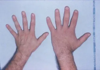
- Acromegaly: too much GH in adults, after the long bones have stopped growing in length: the bones can only thicken, so the hands, feet, lower jaw, nose and brow grow large and coarse. It is usually caused by a benign tumor of the pituitary.
B. Hormones of the neurohypophysis
The posterior lobe stores and releases two hormones made in the hypothalamus, both short peptides of nine amino acids.
- Oxytocin (the “birth hormone”; its drug name is Pitocin): makes the smooth muscle of the uterus contract strongly in labor, which pushes the baby out (parturition); and makes the muscle round the milk glands of the breast contract, squeezing out milk when the baby suckles, the milk-ejection or let-down reflex. In both sexes it is released in moments of closeness and touch, which is why it is nicknamed the “love hormone”; in men it helps the sperm along during ejaculation.
- Vasopressin, or antidiuretic hormone (ADH) (drug name Pitressin): makes the walls of the collecting ducts (and the last part of the distal convoluted tubule) of the kidney’s nephrons let water through, so more water is taken back into the blood and the urine is small and concentrated Grade 12: How urine is formed. Its release is controlled by osmoreceptors in the hypothalamus that sense how concentrated the blood is. In large amounts it also narrows small arteries and raises blood pressure, hence the name vasopressin.
Disorders of ADH:
- Too little: diabetes insipidus. The kidneys cannot keep water, so a person passes huge volumes of dilute urine (5–20 liters a day; diuresis) and is always thirsty. (Insipidus, tasteless: the urine has no sugar, unlike that of diabetes mellitus.)
- Too much: too little urine is made (antidiuresis) and water is kept in the body, diluting the blood.
A boy’s growth hormone is too high from the age of 6. He will most likely develop:
Pick an answer to see why.
Which pair does the posterior pituitary release?
Pick an answer to see why.
A man passes about 10 liters of dilute urine a day and is always thirsty, but his urine has no sugar in it. Which hormone is most likely lacking?
Pick an answer to see why.
Thyroid gland
The thyroid is the largest endocrine gland in the body. It lies in the front of the neck, below the larynx, wrapped round the front and sides of the trachea, level with the fifth cervical to the first thoracic vertebra (C5–T1). It is shaped like a butterfly, or the letter H: two lobes, one each side of the trachea, joined across the front by a narrow band, the isthmus. It weighs 20–25 g, each lobe about 5 cm long. It has a very rich blood supply, so it is darker red than the tissues round it. It is enclosed in a fibrous capsule, and it develops from the endoderm, from the floor of the embryo’s pharynx.
Servier Medical Art (smart.servier.com), CC BY 4.0
Structure under the microscope
The thyroid is made of thousands of round follicles (acini), each a hollow ball lined by a single layer of cuboidal epithelial cells, the follicular or principal cells. The space inside is filled with a sticky, yellowish colloid, made mostly of the protein thyroglobulin, on which the thyroid hormones are made and stored: enough for two or three months. Between the follicles, alone or in small groups, are the parafollicular cells (C cells), which make calcitonin. Capillaries run between the follicles.
Hormones of the thyroid
Under the control of TSH from the pituitary, the follicular cells make triiodothyronine (T₃) and tetraiodothyronine (T₄, thyroxine); the C cells make calcitonin, which is not controlled by TSH. T₃ and T₄ are made by joining iodine to the amino acid tyrosine on thyroglobulin: T₄ carries four iodine atoms, T₃ three. Most of what the gland releases is T₄, but T₃ is the active hormone: in the tissues T₄ loses one iodine atom and becomes T₃.
Functions of T₃ and T₄
- They set the basal metabolic rate (BMR), how fast cells burn food in respiration, and so how much heat the body makes (their calorigenic effect).
- They control the metabolism of carbohydrate, protein and fat.
- They are essential for growth and development, above all of the brain and bones in the unborn baby and in childhood.
- They affect the work of the heart, nerves, muscles and kidney tubules.
- In amphibians they are needed for metamorphosis: a tadpole without thyroid hormone never becomes a frog.
Disorders of T₃ and T₄
1. Hypothyroidism, too little thyroid hormone:
a. Cretinism, from birth (congenital hypothyroidism): stunted growth (a kind of dwarfism, with short limbs), serious intellectual disability because the brain did not develop, undeveloped sex organs, a thick tongue, slow pulse, low blood pressure, low metabolic rate and body temperature. If it is found at birth (many countries test every newborn) and treated with thyroxine tablets at once, the child grows up normal.
b. Myxedema, in adults: a puffy, swollen face and hands (from a jelly-like substance gathering under the skin), dry, pale skin, slow thinking and speech, tiredness, weight gain, feeling cold, low sex drive, and a slow pulse, low blood pressure, low metabolic rate and low temperature.
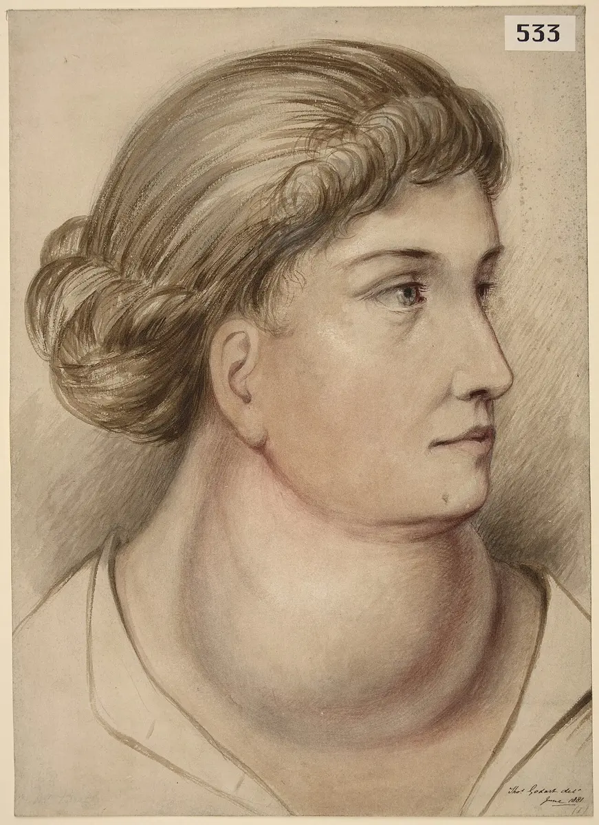
c. Simple goiter: when the diet lacks iodine, the gland cannot make enough hormone; the pituitary keeps sending TSH, and the thyroid swells, its cells multiplying, into a lump in the neck. A goiter shows the gland is struggling; the person may make enough hormone or too little.
d. Hashimoto’s disease (Hashimoto’s thyroiditis): an autoimmune disease in which the body’s own immune system slowly destroys the thyroid, so less and less thyroxine is made. It was first described in 1912 by the Japanese surgeon Hakaru Hashimoto. It is now the commonest cause of hypothyroidism where there is enough iodine.
2. Hyperthyroidism (thyrotoxicosis), too much thyroid hormone:
- Exophthalmic goiter, or Graves’ disease (also Basedow’s or Parry’s disease): an autoimmune disease in which antibodies switch on the TSH receptor, so the thyroid swells and pours out hormone. The signs are bulging eyes (exophthalmos), weight loss despite eating well, restlessness, sleeplessness, sweating, a fast and pounding heartbeat (palpitations), a high metabolic rate and body temperature, and trembling hands.
Calcitonin
Calcitonin, from the C cells, lowers the calcium in the blood when it is too high. It slows the osteoclasts that dissolve bone, so more calcium stays deposited in bone, and it makes the kidneys reabsorb less calcium, so more leaves in the urine. In humans its effect is small: people whose thyroid has been removed keep normal calcium. Its partner and opposite is parathormone.
Which mineral must the diet supply for the thyroid to make thyroxine?
Pick an answer to see why.
A woman has lost weight although she eats more than before, her heart races, her hands tremble and her eyes bulge. She most likely has:
Pick an answer to see why.
A cretin and a pituitary dwarf are both short. What is the main difference?
Pick an answer to see why.
Parathyroid glands
The parathyroid glands are four small, round, yellowish-brown bodies, two on the back of each lobe of the thyroid, partly embedded in it. Each is about 4–6 mm long and weighs about 30–50 mg. Each is wrapped in connective tissue. They develop from the endoderm of the embryo’s pharynx. They are found in amphibians, reptiles, birds and mammals; fishes have none.
Under the microscope they have two kinds of cell: many small, cuboidal chief cells, which make the hormone, and fewer, larger, many-sided oxyphil cells, whose job is unknown; they may be chief cells that have stopped secreting.
Parathormone
The chief cells make parathormone (parathyroid hormone, PTH), also called Collip’s hormone after James Collip, who first extracted it in 1925. PTH keeps the calcium and phosphate of the blood at the right level. When blood calcium falls, PTH raises it again in three ways:
- it releases calcium from bone, by setting the osteoclasts to dissolve bone;
- it makes the kidneys reabsorb more calcium (and get rid of more phosphate);
- it makes the kidneys turn vitamin D into its active form, which makes the intestine absorb more calcium from food.
Calcium is needed for muscle contraction, the clotting of blood, the passing of nerve impulses, and the growth of bones and teeth, so its level is held within narrow limits.
Disorders
- Too little PTH (hypoparathyroidism): blood calcium falls and phosphate rises. Nerves and muscles become over-excitable: tingling and pins and needles round the mouth and in the fingers, muscle cramps and spasms, and in the end tetany, in which muscles, usually of the hands and feet, contract and fail to relax, and convulsions.
- Too much PTH (hyperparathyroidism), usually from a small benign tumor of one gland: calcium is drawn out of bone, so blood calcium rises (hypercalcemia) and more calcium is lost in the urine (hypercalciuria). The bones lose their mineral and grow weak and painful (osteoporosis, with cysts in severe cases), and kidney stones form.
Tetany is caused by:
Pick an answer to see why.
Parathormone and calcitonin are called antagonistic because:
Pick an answer to see why.
Pancreas
The pancreas is a soft, pinkish-yellow, lobed gland about 15 cm long and 60–100 g, lying across the back of the abdomen behind the stomach. Its broad head sits in the C-shaped curve of the duodenum; its body and narrow tail run across to the body’s left, as far as the spleen. It develops from the endoderm of the gut. It is a heterocrine gland: an exocrine gland for digestion and an endocrine gland for blood sugar, in one organ. (Its digestive juice: The pancreas and the intestinal glands.)
Servier Medical Art (smart.servier.com), CC BY 4.0
Structure
About 98–99% of the pancreas is exocrine: lobules of acini, held together by connective tissue. Each acinus is a cluster of secreting cells round a tiny central duct (a pancreatic ductule); they make pancreatic juice, full of digestive enzymes, which flows down the ducts into the duodenum.
Scattered among the acini are 1 to 2 million small groups of endocrine cells, the islets of Langerhans, only about 1–2% of the gland, discovered in 1869 by the German student Paul Langerhans. Each islet is a cluster of a few hundred to a few thousand cells, supplied by nerves of the autonomic nervous system and wrapped in a dense net of capillaries, into which it releases its hormones. Its cells are of four kinds:
| Cell | Share of the islet | Hormone |
|---|---|---|
| Beta (β) cells | about 60% | insulin |
| Alpha (α) cells | about 25% | glucagon |
| Delta (δ) cells | about 10% | somatostatin |
| PP (F) cells | a few | pancreatic polypeptide, which holds back the pancreas’s release of digestive juice |
Servier Medical Art (smart.servier.com), CC BY 4.0
Functions of insulin
Insulin is the body’s only hormone that lowers blood sugar. Released when blood glucose rises after a meal, it:
- makes muscle, fat and other cells take up glucose from the blood and use it in respiration;
- makes the liver and muscles store glucose as glycogen (glycogenesis);
- turns extra glucose into fat;
- speeds up the making of protein and fat, and so, together with growth hormone, is needed for normal growth.
Disorders of insulin
- Diabetes mellitus: too little insulin, or cells that no longer respond to it. Glucose cannot get into the cells, so it piles up in the blood (hyperglycemia) and spills into the urine (glycosuria), taking water with it: the person passes a lot of urine, is always thirsty and hungry, tired, and loses weight. In type 1 diabetes the immune system has destroyed the beta cells, usually in childhood, and the person needs insulin injections for life. In type 2, far commoner and growing fast, the beta cells still make insulin but the body’s cells respond to it poorly (insulin resistance); it comes with age, overweight and too little exercise, and is controlled with diet, exercise and tablets, sometimes insulin.
- Too much insulin lowers blood sugar too far (hypoglycemia): the person shakes, sweats, is confused and may faint or have a fit, because the brain runs on glucose. It happens most often to a diabetic who has injected too much insulin or missed a meal; rarely, a small tumor of the beta cells, an insulinoma, pours out insulin.
Glucagon
Released when blood sugar falls (between meals, in the night), glucagon acts mainly on the liver:
- it breaks stored glycogen down to glucose (glycogenolysis) and releases it into the blood, raising blood sugar;
- it makes the liver take up amino acids and make new glucose from them (gluconeogenesis).
Insulin and glucagon are antagonists, and between them they keep fasting blood sugar at about 70–100 mg per 100 mL. Too little glucagon, which is rare, lets blood sugar fall too low (hypoglycemia).
Somatostatin
Somatostatin from the delta cells holds back the release of insulin and glucagon (and acts on the gut); the somatostatin made by the hypothalamus holds back the release of growth hormone from the pituitary.
Six hours after a meal, which hormone keeps your blood sugar from falling too low?
Pick an answer to see why.
In type 2 diabetes:
Pick an answer to see why.
Adrenal glands
The two adrenal glands (supra-renal glands) sit like caps on top of the kidneys: the right one roughly triangular, the left more like a half-moon. Each is yellowish, about 5 cm long and 4–5 g, has a very rich blood supply, and is wrapped in a thin fibrous capsule.
Servier Medical Art (smart.servier.com), CC BY 4.0
Structure
A lengthwise section (L.S.) shows two quite different parts, of different origin, that work as two separate glands:
- the outer adrenal cortex, about 80–90% of the gland, from the mesoderm; its cells are full of fat droplets and cholesterol, the raw material of its steroid hormones;
- the inner adrenal medulla, from the ectoderm (the neural crest, the same cells that make the sympathetic nerves).
The cortex has three layers, or zones, each making its own kind of hormone; all three kinds are steroids.
| Zone | Where | Makes |
|---|---|---|
| Zona glomerulosa | outer, thin | mineralocorticoids (aldosterone) |
| Zona fasciculata | middle, thickest | glucocorticoids (cortisol) |
| Zona reticularis | inner | sex corticoids (mostly weak androgens) |
The fasciculata and reticularis are controlled by ACTH from the pituitary; the glomerulosa mainly by the salt in the blood (through the kidney’s renin–angiotensin system) and by potassium.
Hormones of the adrenal cortex
a. Mineralocorticoids. The main one is aldosterone. It acts on the distal tubules and collecting ducts of the kidney (and on sweat glands, salivary glands and the gut), making them keep sodium (Na⁺) and get rid of potassium (K⁺). Water follows the sodium, so aldosterone also keeps up the volume of the blood and the blood pressure. It is released when the plasma’s sodium or the blood pressure falls, or its potassium rises.
b. Glucocorticoids: cortisol (the main one, about 95% of their effect), corticosterone and cortisone.
- They control the metabolism of carbohydrate, protein and fat.
- Cortisol makes the liver make new glucose from amino acids (taken from muscle protein) and glycerol (from fat), gluconeogenesis, raising blood sugar.
- It is anti-inflammatory: it calms the immune system and the white blood cells, so cortisol-like drugs (steroids such as prednisolone) are used against inflammation and allergy.
- It is the stress hormone: in illness, injury, hunger or long worry, it keeps energy flowing to the body.
- It helps keep up blood pressure and the normal working of the nervous system.
c. Sex corticoids (gonadocorticoids): small amounts of androgens (male sex hormones) and some estrogen. They help the sex organs and the secondary sexual characters develop and work, in both sexes; in women they are the main source of androgens.
Disorders of the adrenal cortex
- Addison’s disease: the cortex is destroyed (now usually by the body’s own immune system, in Addison’s day mostly by tuberculosis), so too little cortisol and aldosterone are made. Signs: weakness and tiredness, loss of weight, nausea and vomiting, low blood pressure, low Na⁺ and high K⁺ in the plasma, dehydration, low blood sugar, poor resistance to stress and infection, and a bronze darkening of the skin (the pituitary, unchecked, pours out ACTH, which also darkens the skin like MSH).
- Cushing’s syndrome: too much cortisol for a long time, from a tumor or, very often, from long use of steroid medicines. Signs: fat on the trunk and a round moon face, a hump of fat between the shoulders, thin arms and legs from wasted muscle, thin skin with purple stretch marks, slow healing of wounds, high blood sugar, high blood pressure and weak bones. When it is caused by a pituitary tumor making too much ACTH it is called Cushing’s disease; both are named after the American brain surgeon Harvey Cushing, who described it in 1912.
- Hyperaldosteronism (Conn’s syndrome, described by Jerome Conn in 1955): too much aldosterone, usually from a small tumor: the body keeps too much Na⁺ and water and loses K⁺, so blood volume and blood pressure rise, the muscles are weak (low K⁺), and much urine is passed (polyuria).
- Hypoaldosteronism: too little aldosterone: Na⁺ and water are lost in the urine and K⁺ is kept, so the person is dehydrated, with low blood volume and blood pressure.
- Adrenal virilism: too much androgen in a woman (from a tumor, or an inherited fault in making cortisol) gives her male characters: a beard and mustache (hirsutism), a deep voice, more muscle, enlargement of the clitoris, and periods stop.
- Gynecomastia from an adrenal tumor making estrogen in a man: his breasts enlarge, facial hair thins and the voice may rise.
Adrenal medulla
The medulla is made of rounded groups of large, granular chromaffin cells, which are really modified sympathetic nerve cells. They make adrenaline (epinephrine, about 80%) and noradrenaline (norepinephrine, about 20%), together called catecholamines. They are controlled directly by sympathetic nerves, not by the pituitary, so they act within seconds. They prepare the body for the “3 F” response, fight, flight or fright, and so are called emergency hormones; the adrenal gland is the gland of emergency.
Adrenaline:
- is poured out in anger, fear, cold, excitement or pain;
- makes the heart beat faster and harder, raising the cardiac output and the blood pressure;
- narrows the blood vessels of the skin and gut and widens those of the skeletal muscles, sending blood where it is needed to run or fight;
- widens the airways and the pupils;
- raises blood sugar fast by breaking down glycogen (glycogenolysis), and releases fat for fuel;
- is used as a medicine: an injection of adrenaline saves life in a severe allergic reaction (anaphylaxis), and it was once the treatment for asthma attacks.
Noradrenaline acts mainly on the blood vessels: it narrows nearly all of them, keeping the blood pressure up and the circulation normal.
Which hormone is NOT made by the adrenal cortex?
Pick an answer to see why.
A man’s skin has turned bronze, his blood pressure is low, and his plasma has little sodium and too much potassium. Which gland has failed?
Pick an answer to see why.
Why is the adrenal medulla called the gland of emergency?
Pick an answer to see why.
Gonads
The testes and ovaries are the gonads, the primary sex organs. Besides making the gametes (sperm in the testes, eggs in the ovaries) they make the sex steroid hormones, which act on many parts of the body; that is why they are heterocrine glands. (How the gametes are made: Gametogenesis.)
Ovaries, in the pelvis on either side of the uterus, make estrogens and progesterone:
- Estrogens make the uterus and vagina grow and mature; develop the breasts; widen the pelvis; lay fat on the hips, thighs and breasts; build up the lining of the uterus in each menstrual cycle; and bring on the growth of body hair at puberty.
- Progesterone (made mostly by the corpus luteum, and by the placenta in pregnancy) prepares the lining of the uterus to receive an embryo and keeps it in place in pregnancy; it controls the changes of the uterus in the second half of the menstrual cycle; and it develops the milk glands of the breasts in pregnancy.
Testes, in the scrotum, make androgens, the male sex hormones, chiefly testosterone, made by the Leydig (interstitial) cells. (Androgens are also made in small amounts in women, by the ovaries and adrenal glands.) Testosterone develops the male sex organs, before birth and at puberty, and the male secondary sexual characters: more muscle and bone, body and facial hair, broad shoulders, a deeper voice, growth of the penis; and it is needed for making sperm.
Thymus
The thymus lies in the upper front part of the chest, in the mediastinum, behind the sternum and in front of the trachea and heart, below the thyroid, reaching a little into the root of the neck. It is soft, pinkish-gray and lobed. It is relatively large at birth, keeps growing through childhood, and after puberty slowly shrinks (involutes), its tissue replaced by fat, although a little keeps working in adults. Its hormone, thymosin, helps young T lymphocytes multiply and mature, so that they can defend the body.
Pineal gland
The pineal gland is a tiny gland, 0.1–0.2 g, shaped like a pine cone (hence its name), deep in the middle of the brain, between the two cerebral hemispheres at the back of the third ventricle. It makes melatonin, the “sleep hormone”: its release rises in darkness and falls in bright light, so it tells the body when it is night, keeping the sleep–wake cycle (the circadian rhythm) in step with day and night. It may also hold back the development of the sex organs before puberty.
Melatonin is secreted most:
Pick an answer to see why.
Exercise
A. Choose the best alternative
- Which of the following is an endocrine gland?
Pick an answer to see why.
- Who discovered the first hormone, secretin?
Pick an answer to see why.
- What chemical nature do many hormones share with enzymes?
Pick an answer to see why.
- Where is the pituitary gland?
Pick an answer to see why.
- Which hormone is secreted (released) by the posterior lobe of the pituitary gland?
Pick an answer to see why.
- What does antidiuretic hormone do?
Pick an answer to see why.
- Which hormone makes the thyroid gland secrete T₃ and T₄?
Pick an answer to see why.
- Which part of the pituitary gland releases the hormones made by the neurosecretory cells of the hypothalamus?
Pick an answer to see why.
- A patient has a swollen thyroid. Which test is commonly used to find out whether the gland is over-secreting or under-secreting?
Pick an answer to see why.
- What does calcitonin do?
Pick an answer to see why.
- Which disorder results from over-secretion of the thyroid gland?
Pick an answer to see why.
- Which hormone is NOT produced by the adrenal cortex?
Pick an answer to see why.
- What is the main function of cortisol?
Pick an answer to see why.
- Which two minerals does aldosterone balance in the body?
Pick an answer to see why.
- Addison’s disease is caused by a lack of which hormone?
Pick an answer to see why.
- Which condition causes Graves’ disease?
Pick an answer to see why.
B. Answer in brief
What are the differences between exocrine and endocrine glands?
Answer
Which part of the pituitary is known as the storage and release center? Briefly describe its structure and function.
Answer
Describe the causes and symptoms of hypothyroidism and hyperthyroidism.
Answer
Describe the structure and function of the heterocrine gland that lies in the curve of the duodenum.
Answer
What is the function of the parathyroid glands? How do they help keep the body’s calcium in balance?
Answer
Describe briefly the location and the hormonal functions of the gonads.
Answer
What are the differences between enzymes and hormones?
Answer
Describe the structure and function of the adrenal medulla.
Answer
Describe the gland in the brain that keeps the sleep–wake cycle.
Answer
Look at the diagrams and answer the questions.
e. In the drawing below:
Body and glands: BodyParts3D (CC BY 4.0)
- i.Name the parts A to H.
- ii.Which of them is called the gland of emergency?
- iii.What is the function of D?
- iv.Give two differences between B and C.
f. In the drawing below:
Servier Medical Art (smart.servier.com), CC BY 4.0
- i.Name the gland, and say exactly where it is in the body.
- ii.Name the parts a, b and c.
- iii.Write two hormones from part b and two from part c.
Answer
C. Long answer questions
Name the part of the brain that secretes neurohormones and is called the supreme commander of endocrine regulation. Describe its structure and the functions of its secretions.
Answer
Why is the pituitary gland called the master gland? Describe its structure, the functions of its hormones, and its disorders.
Answer
Which gland’s secretion controls the basal metabolic rate? Describe its structure, the functions of its hormones, and its disorders.
Answer
Where are the adrenal glands? Why is the adrenal called the emergency gland? Describe its structure, functions and hormonal disorders.
Answer
Printable PDFs
- Study notes 5 pages
- Worksheet 4 pages Answer key
Quiz yourself: Life Processes and the Human Body, Grades 5–12 →
Practice every question in the book, and keep track of the ones you miss →
A companion to your textbook, not a replacement for it: study from the book your school uses, and where the two differ, follow your teacher. Found a mistake? Please tell us.